
 EN
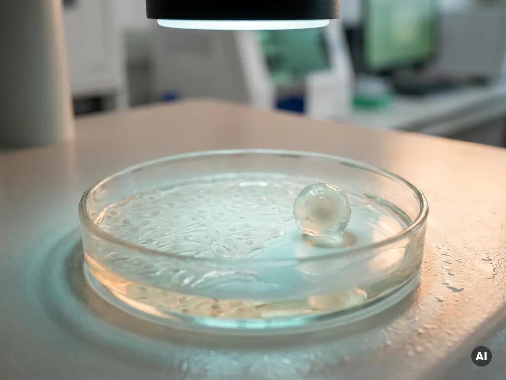
The core difference between 2D and 3D tumor cell culture models lies in how closely they replicate the architecture and biology of real tumors. In 2D models, cancer cells grow as flat monolayers on a plastic surface, while 3D models allow cells to form multilayered structures that better reflect the spatial organization, cell-to-cell interactions, and microenvironmental conditions found in living tissue. For researchers studying tumor biology, drug response, or cancer cell behavior, this structural distinction has significant consequences for experimental outcomes and data relevance. The sections below address the most common questions researchers raise when choosing between these two approaches.
Why do 3D tumor models behave differently from 2D monolayers?
3D tumor models behave differently from 2D monolayers because they recreate the physical and biochemical complexity of the tumor microenvironment, including gradients of oxygen, nutrients, and signaling molecules that are absent in flat culture systems. In a monolayer, every cell has equal access to growth medium and gas exchange, which does not reflect the conditions inside a solid tumor.
In a 3D structure such as a spheroid, cells at the core experience hypoxia and nutrient deprivation, while cells at the periphery remain proliferative. This layered biology drives gene expression patterns, metabolic states, and drug sensitivity profiles that are measurably different from those observed in 2D cell culture. Researchers have found that cells cultured in 3D often display increased resistance to cytotoxic compounds, which may more accurately represent the resistance mechanisms observed in tumor tissue.
Cell-to-cell and cell-to-matrix interactions also play a critical role. In 3D tumor models, cancer cells form physical contacts and communicate through signaling pathways that are structurally constrained in a monolayer. These interactions influence proliferation rates, invasion capacity, and responses to experimental treatments, making 3D systems more physiologically relevant for many research questions. For experiments where the timing of the response is important, real-time cell analysis can complement imaging by continuously monitoring changes in cell behaviour during treatment.
What are the main types of 3D tumor cell culture models?
The main types of 3D tumor cell culture models are spheroids, organoids, scaffold-based cultures, and tumor-on-a-chip systems. Each approach differs in complexity, reproducibility, and the type of tumor biology it can model.
- Spheroids: Self-aggregating clusters of tumor cells grown in non-adherent conditions. They are relatively straightforward to generate and are widely used for drug screening and invasion assays.
- Organoids: More complex 3D structures derived from patient tumor tissue or stem cells, capable of recapitulating organ-specific architecture and genetic heterogeneity. Organoid culture is increasingly used in translational cancer research.
- Scaffold-based models: Cells are seeded into biocompatible matrices such as hydrogels or extracellular matrix-derived materials, which provide structural support and mimic the mechanical properties of tumor stroma.
- Tumor-on-a-chip systems: Microfluidic platforms that integrate 3D cell culture with controlled fluid flow, enabling researchers to study vascularization, immune cell infiltration, and drug perfusion in a dynamic environment.
The choice between these models depends on the research question, the availability of primary tissue, and the degree of biological complexity required. Spheroid culture remains the most accessible entry point, while organoid culture offers greater translational relevance for precision oncology research.
How do 2D and 3D models compare for drug screening accuracy?
3D tumor models generally provide more predictive data for drug screening than 2D cell culture, particularly when assessing compound efficacy, penetration, and resistance mechanisms. Because 2D monolayers expose all cells uniformly to a compound, they tend to overestimate drug sensitivity compared to what is observed in more complex tissue environments.
In 3D systems, drug molecules must diffuse through multiple cell layers to reach the core of a spheroid or organoid. This creates concentration gradients that more closely reflect the pharmacokinetic challenges drugs face in vivo. As a result, compounds that show strong activity in 2D assays may perform differently when tested in 3D tumor models, which can help researchers prioritize candidates more accurately during early-stage screening.
That said, 3D models introduce greater assay variability and require more sophisticated readout technologies. Imaging-based analysis, including high-content imaging and confocal microscopy, is often needed to quantify responses within the full volume of a 3D structure. For large-scale compound screening, researchers in Benelux pharmaceutical and biotech laboratories often combine both approaches, using 2D models for initial high-throughput passes and 3D models for validation and mechanistic studies.
What are the practical limitations of 3D tumor culture systems?
The practical limitations of 3D tumor culture systems include higher technical complexity, reduced throughput, greater cost, and challenges in standardization and reproducibility. These factors can make 3D models more demanding to implement and maintain compared to conventional 2D cell culture.
Generating reproducible spheroids or organoids requires careful control of seeding density, matrix composition, and culture conditions. Variability in size and morphology between batches can introduce noise into experimental data. Organoid culture in particular demands specialized media formulations and expertise in handling primary tissue, which increases the barrier to entry for laboratories without prior experience.
Imaging and quantification also present challenges. Standard plate readers and bright-field microscopy are often insufficient for capturing the full three-dimensional biology of these models. Researchers typically require confocal imaging, light-sheet microscopy, or dedicated 3D analysis software to extract meaningful data from intact structures. Confocal and widefield imaging, combined with Z-stack acquisition and automated image analysis, can help you quantify spheroid morphology, viability, growth and treatment response.
Finally, throughput remains a constraint. While miniaturized formats and automation are improving this, 3D systems are generally not yet as amenable to ultra-high-throughput screening as optimized 2D assays. Laboratories must weigh the biological relevance gained against the operational investment required.
When should researchers choose 2D over 3D tumor models?
You should choose 2D tumor cell culture models when speed, throughput, and simplicity are priorities, or when the experimental question does not require three-dimensional tissue architecture. 2D monolayers remain highly appropriate for many standard applications in cancer research.
Suitable scenarios for 2D cell culture include:
- Initial compound screening: When evaluating large libraries of compounds for basic cytotoxicity or growth inhibition, 2D models offer the throughput and assay consistency needed for rapid prioritization.
- Genetic and molecular studies: Transfection, gene editing, and Western blot-based analyses are often more straightforward in adherent monolayers, where cell access and lysis are uncomplicated.
- Proliferation and cell cycle assays: Standard growth kinetics, cell cycle profiling, and apoptosis assays are well established and validated in 2D formats.
- Protocol development and optimization: When developing a new assay or standardizing a workflow, 2D models provide a simpler baseline before transitioning to more complex systems.
- Resource-limited settings: Where budget, equipment, or expertise constrains the use of 3D systems, 2D models continue to generate valuable mechanistic data.
The key principle is to match the model to the question. 2D cell culture is not obsolete; it remains an essential tool in the cancer researcher’s toolkit when used for the right applications.
What instrumentation supports 2D and 3D tumor cell culture workflows?
Both 2D and 3D tumor cell culture workflows rely on a range of laboratory instruments, from basic cell maintenance equipment to advanced imaging and analysis platforms. The instrumentation required becomes more specialized as the complexity of the culture model increases.
For 2D cell culture, core instruments include CO2 incubators, biosafety cabinets, inverted microscopes, and multimode plate readers capable of measuring absorbance, fluorescence, and luminescence. These tools support routine cell maintenance, viability assays, and endpoint measurements.
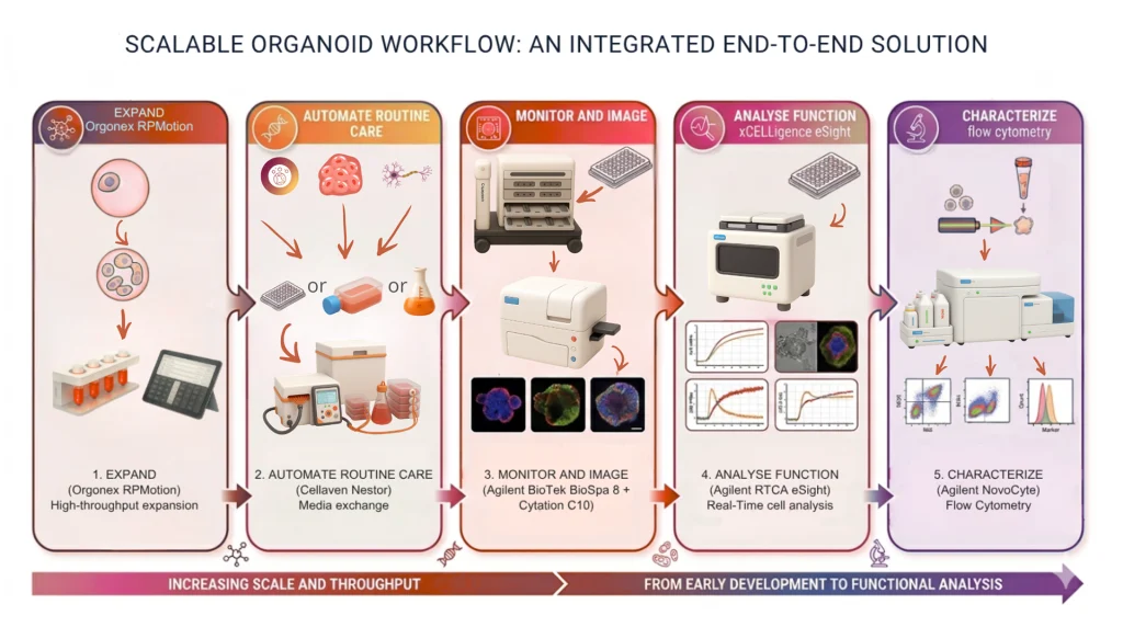
For 3D tumor models, additional or more advanced instrumentation is typically required. High-content imaging systems can capture spatial information across the full volume of a spheroid, while confocal and light-sheet microscopes enable detailed structural analysis. Liquid handling systems improve the consistency of spheroid seeding and medium exchange, which is critical for reproducibility. Automated cell culture platforms can reduce manual handling variability, particularly when working with sensitive organoid cultures over extended periods.
Multimode readers with z-stack imaging capability and dedicated 3D analysis software are increasingly valuable for quantifying dose-response relationships within intact 3D structures. For researchers scaling up organoid workflows, bioreactor-based systems offer controlled environments for longer-term culture and expansion.
How BioSPX supports 2D and 3D tumor cell culture research
BioSPX offers a curated portfolio of laboratory instruments tailored to the needs of cancer researchers and life science laboratories across Belgium, the Netherlands, and Luxembourg. Whether you are working with standard 2D monolayer assays or transitioning to more complex 3D tumor models, BioSPX provides the instrumentation and expert guidance to support your workflow.
- Multimode readers and high-content imaging systems for quantitative analysis of both 2D and 3D cultures
- Liquid handling solutions to improve seeding consistency and medium exchange reproducibility
- Automated cell culture platforms, including solutions for organoid and 3D model maintenance
- Bioreactor systems designed for organoid research and extended 3D culture workflows
- Installation, user training, and application support from product specialists with deep domain expertise
If you are evaluating which instruments best fit your tumor cell culture research, contact BioSPX to speak with a specialist who can advise on the right configuration for your laboratory.
Frequently Asked Questions
How do I transition my lab from 2D to 3D tumor cell culture without disrupting existing workflows?
The most practical approach is to run 2D and 3D models in parallel during an initial transition period, allowing you to validate your 3D results against your established 2D baseline before fully committing to the new format. Start with a well-characterized cell line and a straightforward spheroid format, such as ultra-low attachment plates, before moving to more complex scaffold-based or organoid systems. Investing in training and protocol standardization early — particularly around seeding density and batch consistency — will significantly reduce variability and shorten the learning curve.
Can patient-derived tumor cells be used in both 2D and 3D culture systems, and which performs better?
Patient-derived tumor cells can be used in both formats, but they tend to perform more predictably and retain greater clinical relevance in 3D systems, particularly organoid culture. In 2D monolayers, primary tumor cells often lose their original phenotype over successive passages due to selection pressure from the plastic surface and uniform nutrient exposure. Organoids derived from patient tissue better preserve the genetic heterogeneity and histological characteristics of the original tumor, making them especially valuable for translational research and personalized medicine applications.
What are the most common mistakes researchers make when setting up spheroid cultures for the first time?
The most frequent mistakes include inconsistent seeding density, which leads to highly variable spheroid sizes that compromise assay reproducibility, and using standard tissue culture-treated plates instead of non-adherent or ultra-low attachment surfaces. Researchers also commonly underestimate the importance of medium exchange timing — disturbing early aggregates before they have consolidated can disrupt spheroid formation entirely. Establishing a strict, documented protocol for each cell line, including optimal seeding density and the earliest safe time point for medium changes, is essential before moving into experimental work.
How should drug concentration ranges be adjusted when moving from 2D to 3D tumor models?
When transitioning drug testing from 2D to 3D models, researchers should generally expect to test higher concentration ranges, as 3D structures frequently exhibit increased drug resistance due to diffusion barriers, hypoxic cores, and enhanced cell-to-matrix interactions. A practical starting point is to extend your existing 2D concentration range by at least one to two orders of magnitude on the higher end and perform a full dose-response curve rather than relying on single-point measurements. This recalibration is particularly important for compounds targeting proliferating cells, since the quiescent cell populations in spheroid cores may respond very differently than the actively dividing cells your 2D IC50 data reflects.
Are there specific cell lines that are better suited to 3D tumor culture than others?
Yes — not all cell lines form well-defined 3D structures with equal efficiency, and this is one of the practical realities researchers encounter early in the transition. Cell lines with strong homotypic adhesion properties, such as many breast, colorectal, and lung cancer lines including MCF-7, HCT116, and A549, tend to form compact, reproducible spheroids under standard non-adherent conditions. Other lines, particularly those with low E-cadherin expression, may require a scaffold-based matrix such as Matrigel or a collagen hydrogel to maintain structural integrity. Checking published literature for your specific cell line before committing to a culture format will save considerable time and resources.
What viability assays are compatible with 3D tumor models, and how do they differ from standard 2D assays?
Luminescence-based viability assays, such as CellTiter-Glo 3D, are among the most widely used and reliable options for 3D tumor models because they lyse the entire structure and measure total ATP content, providing a whole-spheroid readout without the need for imaging. Standard colorimetric assays like MTT are generally less suitable for intact 3D structures because the dye penetration and formazan solubilization steps are inconsistent across the depth of the spheroid. For researchers who want spatial information — for example, distinguishing live and dead cell zones within a spheroid — fluorescent dye combinations imaged by confocal microscopy offer greater resolution but require more advanced instrumentation and analysis workflows.
How is artificial intelligence or image analysis software changing the way researchers quantify 3D tumor model data?
AI-assisted image analysis is rapidly improving the speed and objectivity of 3D tumor model quantification, particularly for tasks like automated spheroid segmentation, volume measurement, and classification of morphological changes across large image datasets. Platforms integrated into high-content imaging systems can now detect subtle phenotypic shifts — such as changes in spheroid compactness or invasion front geometry — that would be impractical to score manually at scale. For laboratories scaling up 3D drug screening workflows, these tools significantly reduce the analytical bottleneck that has historically made 3D assays less amenable to higher-throughput applications.
Related Articles
- How does sample preparation affect spatial protein analysis?
- How does photobleaching affect fluorescence-based molecular interaction measurements?
- Why is proliferation kinetics more informative than a single growth measurement?
- What can continuous cell monitoring reveal that endpoint assays cannot?
- What is the difference between curative and palliative cancer treatment?
This content was generated with the help of AI and it may contain mistakes