
 EN
The most significant advances in oncology in 2026 centre on three converging forces: artificial intelligence accelerating discovery, liquid biopsy enabling earlier detection, and increasingly precise immunotherapy approaches reshaping how cancer research is conducted. These developments are not isolated breakthroughs but interconnected shifts that are collectively transforming the scientific landscape. The sections below unpack each of these advances and the questions researchers are asking about them.
Which cancer treatments made the biggest breakthroughs in 2026?
The most notable cancer treatment breakthroughs in 2026 involve next-generation antibody-drug conjugates, CAR-T cell refinements, and combination immunotherapy regimens that are being investigated for a wider range of tumour types. These approaches share a common thread: they aim to act with greater specificity, reducing off-target effects while increasing biological impact at the tumour site.
Antibody-drug conjugates, which link a targeting antibody to a cytotoxic payload, have advanced significantly in terms of linker stability and payload diversity. Researchers are exploring how these constructs can be optimised for solid tumours, which historically have been more resistant to such approaches than haematological cancers.
CAR-T cell therapies have also seen meaningful refinements in 2026, particularly in manufacturing efficiency and the development of allogeneic (off-the-shelf) formats. These formats are being investigated for their potential to make cell therapy research more scalable and reproducible, which is a critical consideration for laboratory teams working in the Benelux region and beyond.
How has AI changed oncology diagnosis and drug development in 2026?
AI has changed oncology in 2026 by accelerating target identification, improving the analysis of complex multi-omics datasets, and enabling more accurate pattern recognition in imaging data. These capabilities are allowing research teams to move from hypothesis to experimental validation faster than was previously possible.
In drug development workflows, machine learning models are being used to predict molecular interactions, prioritise candidate compounds, and identify potential resistance mechanisms before they emerge in experimental settings. This is particularly valuable in early-stage oncology research, where the search space for viable targets is enormous.
In diagnostic research, AI-assisted image analysis is being applied to histopathology and radiology data to identify morphological features that may be difficult for human observers to detect consistently. While these tools are research instruments rather than clinical decision-makers, they are generating hypotheses that drive further experimental investigation.
What role does liquid biopsy play in cancer detection in 2026?
Liquid biopsy plays an increasingly central role in cancer detection research in 2026 by enabling the analysis of circulating tumour DNA, RNA, proteins, and extracellular vesicles from blood or other biofluids. This approach is being investigated as a way to detect tumour-derived signals earlier and with less invasive sampling than traditional tissue biopsy.
The key scientific interest in liquid biopsy lies in its potential for longitudinal monitoring. Because samples can be collected repeatedly over time, researchers can study how tumour-associated biomarkers evolve in response to experimental interventions. This makes it a valuable tool for understanding resistance mechanisms and disease dynamics.
In 2026, advances in sequencing sensitivity and signal enrichment methods have improved the ability to detect low-abundance circulating tumour DNA. Multi-analyte panels that combine several biomarker types are also being developed to increase the specificity of detection signals, which is a key challenge in this field.
How is personalised medicine reshaping cancer care in 2026?
Personalised medicine is reshaping cancer research in 2026 by shifting the focus from tumour type to tumour biology, meaning that molecular profiling rather than organ of origin increasingly guides how experimental therapies are designed and tested. This approach, often called precision oncology, relies on identifying actionable alterations in individual tumour genomes.
Multi-omics profiling, which integrates genomic, transcriptomic, proteomic, and epigenomic data, is becoming more accessible to research laboratories. The ability to layer these data types gives researchers a more complete picture of tumour heterogeneity and the signalling pathways that sustain cancer cell growth.
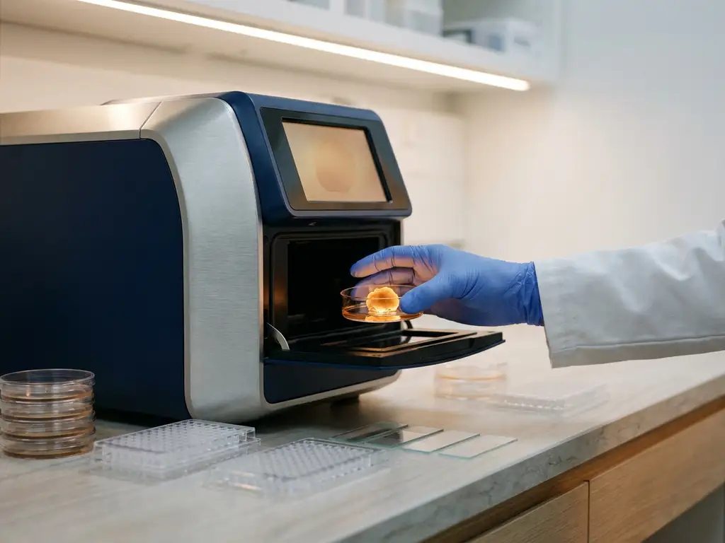
Organoid technology is playing a growing role in this space. Patient-derived organoids, which are three-dimensional tumour models grown from biopsy material, allow researchers to test experimental compounds in a system that preserves much of the original tumour’s biological complexity. This is an area where laboratory instrumentation for organoid culture and analysis is directly enabling precision oncology research workflows. High-content imaging can help researchers characterise how patient-derived organoids respond to experimental treatments. The Cytation C10 combines advanced imaging and quantitative analysis to support the study of 3D cell models, enabling researchers to assess changes in cell morphology, viability, and treatment response within more physiologically relevant experimental systems.
What are the most promising immunotherapy developments in 2026?
The most promising immunotherapy developments in 2026 include bispecific T-cell engagers, tumour microenvironment-targeting strategies, and personalised neoantigen vaccine approaches. Each of these is being investigated for its potential to overcome the immune evasion mechanisms that limit the effectiveness of earlier immunotherapy generations.
Real-time cell analysis can help researchers quantify the effects of emerging immunotherapies directly in cell-based assays. The xCELLigence RTCA platform enables label-free, real-time monitoring of cellular responses, including immune-cell-mediated tumour cell killing, making it useful for evaluating approaches such as bispecific T-cell engagers, checkpoint inhibitors, and engineered T-cell therapies.
Research interest in the tumour microenvironment has grown substantially. Scientists are investigating how immunosuppressive cell populations, such as regulatory T cells and myeloid-derived suppressor cells, create barriers to immune activation within the tumour. Understanding these dynamics at the molecular level is considered essential for designing the next generation of combination approaches. Multiparameter flow cytometry provides a way to characterise the immune cell populations involved in the tumour microenvironment. NovoCyte flow cytometers enable researchers to analyse multiple cellular markers simultaneously, supporting detailed profiling of immune-cell phenotypes and responses during cancer immunotherapy research.
Neoantigen-based vaccine research is also advancing, with improved methods for identifying tumour-specific mutations that can serve as immunological targets. The computational and laboratory workflows required to design and validate these candidates are becoming more refined, making this an active area of methodological development in 2026.
What challenges still limit oncology progress despite 2026 advances?
Despite the advances made in oncology in 2026, several fundamental challenges remain. These include tumour heterogeneity, acquired resistance to targeted approaches, the complexity of the tumour microenvironment, and the reproducibility of experimental models. No single breakthrough has resolved these issues, and they continue to shape research priorities.
Key ongoing challenges include:
- Tumour heterogeneity: Cancer cells within a single tumour can carry different mutations, making uniform targeting difficult to achieve in experimental models
- Resistance mechanisms: Tumours can adapt to selective pressure from targeted interventions, developing new survival pathways that require further investigation
- Model fidelity: Standard two-dimensional cell culture models do not fully replicate the three-dimensional architecture and microenvironmental conditions of actual tumours
- Data integration: Combining multi-omics datasets into actionable biological insights remains computationally and analytically demanding
- Reproducibility: Ensuring that experimental results are consistent across laboratories and conditions is a persistent challenge in cancer research
Addressing these challenges requires not only scientific innovation but also improvements in the tools and platforms used to conduct research. Better experimental models, more sensitive detection instruments, and more automated workflows all contribute to closing the gap between biological insight and reliable, reproducible data.
How BioSPX supports oncology research in the Benelux
Research teams working on oncology advances in Belgium, the Netherlands, and Luxembourg need instruments and workflows that match the complexity and precision demands of modern cancer research. BioSPX supports these laboratories with a portfolio of life science instruments and expert guidance tailored to the Benelux research environment.
- Organoid research tools: Solutions such as bioreactor systems support the three-dimensional tumour models increasingly used in precision oncology workflows
- Cell culture automation: Automated cell culture systems reduce manual variability and free researchers to focus on experimental design rather than routine maintenance tasks
- Imaging and microscopy: High-content imaging systems support the visual analysis of cellular responses in complex tumour models
- Liquid handling: Precise liquid handling platforms improve reproducibility in assay preparation and multi-sample workflows
- Expert support: Product specialists advise on instrument configuration and experimental setup, with a service department ensuring correct installation and rapid response when needed
Whether your laboratory is exploring immunotherapy mechanisms, validating liquid biopsy assays, or building organoid-based research models, the right instrumentation makes a measurable difference. Contact BioSPX to discuss which solutions best fit your oncology research goals.
Frequently Asked Questions
How can a research lab get started with liquid biopsy workflows if they have no prior experience in this area?
The most practical starting point is to focus on a single analyte type — circulating tumour DNA is the most established — before expanding to multi-analyte panels. Labs should assess their existing sequencing infrastructure and identify any sensitivity gaps, as low-abundance ctDNA detection demands high-performance library preparation and enrichment protocols. Partnering with an instrument specialist who understands the specific sensitivity and throughput requirements of liquid biopsy can help avoid costly mismatches between workflow needs and platform capabilities.
What are the most common mistakes researchers make when working with patient-derived organoids?
One of the most frequent pitfalls is insufficient quality control during the establishment phase — organoids that drift from their original tumour biology over successive passages can produce misleading experimental data. Inconsistent extracellular matrix composition and manual handling variability are also major sources of irreproducibility between experiments and between laboratories. Investing in automated culture systems and standardised imaging-based viability assessments from the outset significantly improves the reliability of downstream results.
How do researchers ensure reproducibility when integrating multi-omics datasets from different platforms?
Reproducibility in multi-omics integration depends heavily on standardising sample preparation protocols upstream, since batch effects introduced at the wet lab stage are difficult to correct computationally after the fact. Using validated bioinformatics pipelines with clearly documented normalisation steps, and where possible benchmarking against publicly available reference datasets, adds an important layer of analytical consistency. Many research teams are also adopting LIMS (Laboratory Information Management Systems) to maintain traceability across sample types and experimental runs.
Can smaller or mid-sized Benelux research labs realistically implement AI-assisted image analysis, or is this only feasible for large institutions?
AI-assisted image analysis is increasingly accessible to labs of all sizes, particularly as cloud-based analysis platforms reduce the need for on-site computational infrastructure. Many high-content imaging systems now come with integrated AI modules that can be configured without specialist data science expertise, making entry-level implementation achievable for focused applications such as cell morphology scoring or organoid size quantification. The key is to match the analytical ambition to the available training data — AI models perform poorly when trained on small or poorly annotated image sets.
What should oncology research teams prioritise when evaluating new instrumentation for CAR-T or other cell therapy workflows?
The most critical factors are process consistency and scalability — instruments that introduce variability at the cell expansion or formulation stage can compromise the biological relevance of experimental results. Teams should also evaluate the instrument’s compatibility with closed-system processing, which reduces contamination risk and is increasingly important for allogeneic CAR-T research formats. Asking vendors for application-specific validation data and requesting a demonstration with your own cell types before committing to a platform is strongly advisable.
How is the tumour microenvironment typically modelled in vitro, and what are the current limitations of these models?
The most common in vitro approaches include co-culture systems that combine tumour cells with immune or stromal cell populations, and three-dimensional organoid or spheroid models that better replicate spatial architecture than standard 2D monolayers. However, even advanced 3D models struggle to fully recapitulate the dynamic vascularisation, oxygen gradients, and immune cell trafficking that characterise the in vivo tumour microenvironment. Microfluidic tumour-on-a-chip platforms are an emerging solution to some of these limitations, though they currently require specialised expertise and are not yet widely standardised across laboratories.
What practical steps can a lab take today to improve the reproducibility of its oncology experiments?
The single highest-impact step most labs can take is to audit and standardise their cell culture practices, including passage number tracking, mycoplasma testing schedules, and media preparation protocols, as these upstream variables account for a disproportionate share of irreproducible results. Introducing automated liquid handling for assay preparation reduces pipetting variability and is particularly valuable in high-throughput screening or multi-sample biomarker workflows. Documenting experimental conditions in sufficient detail to allow independent replication — including instrument settings, reagent lot numbers, and environmental parameters — is also essential and often underestimated.
Related Articles
- What are the challenges of multiplexed molecular analysis in tissue samples?
- How does FLIM-FRET preserve spatial information that bulk assays lose?
- What causes genomic instability in cancer cells?
- What is the difference between benign and malignant tumors?
- What is the difference between oncology and hematology?
This content was generated with the help of AI and it may contain mistakes