
 EN
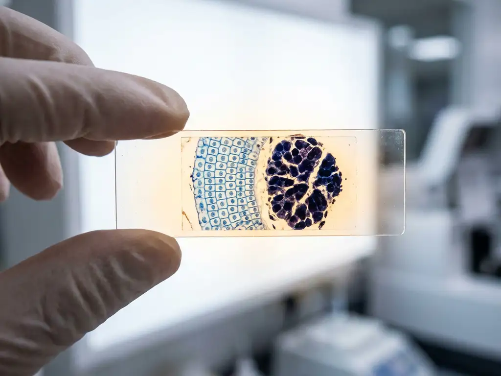
A benign tumor is a non-cancerous growth that does not invade surrounding tissue or spread to other parts of the body. A malignant tumor, by contrast, is cancerous: it grows aggressively, invades neighboring tissues, and can metastasize through the bloodstream or lymphatic system. Understanding the difference between benign and malignant tumors is fundamental to oncology research, diagnostic pathology, and the development of new detection methods. The sections below address the most common questions researchers and life science professionals encounter when working with tumor biology.
Can a benign tumor become malignant over time?
Yes, some benign tumors can transform into malignant ones, though this does not happen with all tumor types. This process, called malignant transformation, occurs when cells within a benign growth accumulate additional genetic mutations that disrupt normal growth regulation. The likelihood and timeline of transformation vary considerably depending on the tissue of origin and the specific cellular characteristics involved.
Certain benign lesions are considered precancerous because they carry a measurably higher risk of progression. Adenomatous polyps in the colon, for example, are well-studied in research contexts as models for stepwise tumor progression. Similarly, some forms of dysplasia, where cells show abnormal but non-invasive changes, are investigated as intermediate states between benign and malignant biology.
Not all benign tumors carry this risk. Lipomas, for instance, rarely, if ever, undergo malignant transformation. Research into which tumor types are prone to progression, and under what molecular conditions, remains an active area within cancer biology and translational research.
How do benign and malignant tumors grow differently?
Benign tumors grow slowly, remain localized, and are typically surrounded by a fibrous capsule that keeps them contained. Malignant tumors grow more rapidly, lack clear boundaries, and invade surrounding tissue by breaking through the basement membrane and infiltrating adjacent structures. This invasive growth pattern is one of the defining biological features that distinguishes malignancy from benign disease.
At the cellular level, malignant tumors show greater genetic instability, higher rates of cell division, and impaired apoptosis, meaning cells fail to undergo normal programmed death. Benign tumor cells generally retain a more normal appearance and function closer to the tissue they originated from.
Another critical difference is metastatic potential. Benign tumors do not metastasize. Malignant tumors can shed cells into the bloodstream or lymphatic system, establishing secondary tumors at distant sites. This capacity for metastasis is what makes malignant disease significantly more complex to study and address in research settings.
What causes a tumor to become malignant?
Malignant transformation is driven by the accumulation of genetic and epigenetic changes in cells. Key drivers include mutations in proto-oncogenes, which promote cell growth, and tumor suppressor genes, which normally act as brakes on cell division. When both types of regulatory mechanisms are compromised, cells can proliferate uncontrollably and acquire invasive properties.
Several factors are investigated in research for their role in promoting these mutations:
- Chronic inflammation, which creates a microenvironment conducive to DNA damage
- Exposure to carcinogens, including certain chemicals and types of radiation
- Viral infection, as some viruses integrate into the host genome and disrupt normal cell regulation
- Inherited genetic predispositions that reduce the cell’s ability to repair DNA errors
- Epigenetic alterations that silence tumor suppressor genes without changing the DNA sequence itself
Research in this area focuses heavily on the tumor microenvironment, the surrounding network of immune cells, signaling molecules, and structural proteins that can either suppress or facilitate malignant progression.
How are benign and malignant tumors diagnosed?
Tumor diagnosis relies on a combination of imaging, histopathological analysis, and molecular profiling. Histopathology remains the gold standard: a tissue biopsy is examined under a microscope to assess cell morphology, growth patterns, and the presence of invasive features. Malignant tumors typically show nuclear atypia, irregular mitotic figures, and loss of normal tissue architecture.
In research and clinical laboratory settings, several diagnostic approaches are used in parallel:
- Imaging techniques such as MRI, CT, and ultrasound provide structural information about tumor size, location, and boundaries
- Immunohistochemistry uses antibody-based staining to identify specific protein markers associated with malignancy
- Molecular diagnostics, including genomic sequencing, identify mutations and expression profiles that characterize tumor type and behavior
- Flow cytometry and cell imaging are used in research workflows to analyze cell populations and detect abnormal phenotypes
- Liquid biopsy methods are being investigated for their ability to detect circulating tumor DNA as a non-invasive diagnostic approach
Advances in imaging and molecular biology instrumentation have significantly improved the resolution and accuracy with which researchers can characterize tumor tissue, supporting more precise classification between benign and malignant growths.
What are the treatment differences for benign versus malignant tumors?
In research contexts, the distinction between benign and malignant tumors shapes the experimental models, assays, and intervention strategies under investigation. Benign tumors are primarily studied for their potential to transform, their structural biology, or as controls in comparative experiments. Malignant tumors are investigated for mechanisms of invasion, metastasis, drug resistance, and therapeutic targeting.
From a scientific methodology perspective, malignant tumor research involves considerably more complex experimental systems. Three-dimensional cell culture models, organoid platforms, and co-culture systems that replicate the tumor microenvironment are increasingly used to study malignant behavior under conditions that more closely resemble in vivo biology. These approaches are being explored for their value in modeling tumor progression and evaluating potential intervention strategies.
Benign tumor research, while less prominent, contributes to understanding baseline tissue biology and the early molecular events that may precede malignant transformation.
Which tumor types are most commonly misclassified as benign or malignant?
Several tumor types present diagnostic challenges because their histological features overlap between benign and malignant categories. Misclassification in research and pathology contexts can occur when tumors show intermediate characteristics that do not fit neatly into either category.
Commonly ambiguous tumor types include:
- Borderline ovarian tumors, which show some features of malignancy but lack clear stromal invasion
- Gastrointestinal stromal tumors (GISTs), where risk stratification between benign and malignant behavior depends on size and mitotic rate rather than histology alone
- Phyllodes tumors of the breast, which exist on a spectrum from benign to malignant and require careful molecular analysis
- Well-differentiated liposarcoma, which can closely resemble a benign lipoma under standard microscopy
- Atypical meningiomas, which occupy an intermediate grade between clearly benign and clearly malignant brain tumors
Advances in molecular profiling and high-resolution imaging are helping researchers develop more reliable classification criteria for these ambiguous cases. In Benelux research institutions and biotech environments, improved instrumentation for cell imaging and molecular analysis is contributing to more accurate tumor characterization at the research level.
How BioSPX supports tumor biology research in the Benelux
Understanding the biological differences between benign and malignant tumors requires precise, reliable instrumentation. Research teams across Belgium, the Netherlands, and Luxembourg rely on advanced tools for imaging, cell analysis, and experimental modeling to investigate tumor behavior at the cellular and molecular levels.
BioSPX supports life science researchers in the Benelux region with a portfolio of instruments suited to tumor biology workflows, including:
- Multimode readers and imaging systems for high-resolution cell and tissue analysis
- Liquid handling solutions that improve reproducibility in complex assay workflows
- Organoid research platforms that enable three-dimensional tumor modeling
- Automation systems that reduce manual variability in cell culture and experimental preparation
BioSPX’s product specialists advise on instrument selection and experimental setup, while the service team ensures correct installation and ongoing support. To discuss which instruments best fit your tumor research workflow, contact the BioSPX team directly.
Frequently Asked Questions
What in vitro models are best suited for studying malignant tumor behavior in a research setting?
Three-dimensional culture systems, including organoids and spheroids, are increasingly preferred over traditional 2D monolayer cultures because they better replicate the spatial architecture, oxygen gradients, and cell-cell interactions found in vivo. Co-culture systems that incorporate stromal and immune cells are particularly valuable for studying invasion and tumor microenvironment dynamics. The choice of model ultimately depends on the research question — organoid platforms are well-suited for studying tumor progression and drug response, while transwell invasion assays remain practical for assessing malignant cell behavior in a more controlled format.
What molecular markers are commonly used to distinguish malignant from benign tumor cells in research workflows?
Several protein and genomic markers are routinely used to characterize malignancy, including Ki-67 as an indicator of proliferative activity, p53 mutation status as a marker of genomic instability, and E-cadherin loss as a sign of epithelial-to-mesenchymal transition. Immunohistochemistry panels targeting these and other markers — such as HER2, EGFR, or vimentin — are commonly applied in research workflows to build a molecular profile of the tumor. Genomic sequencing adds another layer by identifying specific driver mutations that define tumor type and behavior beyond what histology alone can reveal.
How should researchers handle ambiguous tumor samples that don't clearly fall into benign or malignant categories?
When histological features are inconclusive, combining multiple diagnostic modalities is the most reliable approach — integrating immunohistochemical staining, molecular profiling, and high-resolution imaging rather than relying on morphology alone. For tumor types known to occupy a biological spectrum, such as phyllodes tumors or GISTs, risk stratification criteria specific to that tumor type should guide classification. Consulting current pathology consensus guidelines and, where possible, applying genomic sequencing to identify known malignancy-associated mutations can significantly reduce misclassification risk in research contexts.
What are the most common experimental mistakes when working with malignant tumor cell lines in the lab?
One of the most frequent issues is failing to authenticate cell lines before use, which can lead to misidentified or cross-contaminated cultures that compromise the validity of results — STR profiling is the standard method to verify cell line identity. Researchers also commonly overlook the impact of passage number, as highly passaged malignant cell lines can drift significantly from the original tumor’s molecular characteristics. Additionally, using 2D monoculture as the sole experimental system can produce results that do not translate well to in vivo conditions, particularly when studying invasion or drug resistance mechanisms that are strongly influenced by the tumor microenvironment.
How is liquid biopsy being used in tumor research, and what are its current limitations?
Liquid biopsy — primarily the detection and analysis of circulating tumor DNA (ctDNA) in blood — is being investigated as a minimally invasive method for tumor characterization, treatment monitoring, and early detection of malignant disease. In research settings, it offers the advantage of capturing tumor heterogeneity across multiple sites without requiring surgical tissue sampling. However, current limitations include low ctDNA abundance in early-stage or benign-adjacent tumors, sensitivity challenges with certain tumor types, and the need for highly standardized pre-analytical protocols to ensure reproducible results across studies.
Are there specific biosafety or containment considerations when working with malignant tumor samples or cell lines?
Malignant human tumor cell lines are typically handled under Biosafety Level 2 (BSL-2) conditions, which require appropriate personal protective equipment, work within a biological safety cabinet, and strict waste decontamination procedures. Researchers should also be aware of any viral components associated with specific tumor lines — for example, cell lines derived from virus-associated tumors may carry additional biosafety considerations. Institutional biosafety committees should be consulted when establishing new tumor research workflows, particularly when working with primary patient-derived material or novel organoid systems.
How can research teams in the Benelux access instrument demonstrations or application support for tumor biology workflows?
BioSPX offers direct consultation with product specialists who can advise on instrument selection based on your specific tumor research workflow — whether that involves high-content imaging, organoid culture, liquid handling automation, or molecular analysis. Teams interested in evaluating instruments before purchase can reach out to discuss demonstration options or application-specific guidance. The BioSPX service team also provides installation support and ongoing technical assistance to ensure instruments perform reliably within established research protocols.
Related Articles
- Why is proliferation kinetics more informative than a single growth measurement?
- What is the tumor microenvironment and why does it matter in research?
- How do oncogenes differ from tumor suppressor genes in cancer development?
- What is the difference between apoptosis and necrosis in cancer cells?
- How do oncologists decide which cancer treatment to recommend?
This content was generated with the help of AI and it may contain mistakes