
 EN
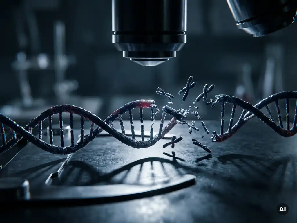
Genomic instability in cancer cells is caused by a combination of defective DNA repair mechanisms, replication errors, environmental mutagens, and dysfunctional cell cycle checkpoints. These disruptions allow mutations and chromosomal abnormalities to accumulate over successive cell divisions, driving the genetic diversity that enables tumors to grow, adapt, and resist treatment. The sections below unpack the key mechanisms and research questions behind this process.
How does genomic instability drive tumor progression?
Genomic instability accelerates tumor progression by generating a continuous supply of genetic variation within a tumor cell population. This variation acts as raw material for natural selection: cells that acquire mutations conferring growth advantages, resistance to cell death, or the ability to invade surrounding tissue are selected for, while others are outcompeted. The result is a progressively more aggressive tumor over time.
Researchers studying tumor evolution increasingly recognize that genomic instability is not merely a byproduct of cancer but a core engine of disease progression. A genetically heterogeneous tumor contains subpopulations of cells with distinct mutation profiles, which complicates research into therapeutic resistance and disease modeling. Understanding the dynamics of this instability is central to cancer biology research across laboratories in Belgium, the Netherlands, and Luxembourg.
What are the main types of genomic instability in cancer?
The main types of genomic instability in cancer cells are chromosomal instability, microsatellite instability, and nucleotide instability. Each operates through a distinct mechanism and leaves a characteristic molecular fingerprint that researchers can detect and study in tumor samples.
- Chromosomal instability (CIN): The most common form, involving gains, losses, or rearrangements of whole chromosomes or large chromosomal segments. CIN arises from errors during cell division, particularly at the mitotic spindle checkpoint.
- Microsatellite instability (MSI): Caused by defective mismatch repair, MSI results in the expansion or contraction of short repetitive DNA sequences known as microsatellites. It is frequently observed in colorectal and endometrial cancers.
- Nucleotide instability: Involves point mutations and small insertions or deletions caused by errors in base excision repair or nucleotide excision repair pathways.
Each type of instability produces a distinct mutational landscape, and researchers use sequencing technologies and genomic profiling tools to characterize these patterns in cancer models and primary tumor samples.
What causes DNA repair mechanisms to fail in cancer cells?
DNA repair mechanisms fail in cancer cells primarily because of mutations or epigenetic silencing in the genes that encode repair proteins. When these genes are inactivated, the cell loses its ability to detect and correct DNA damage accurately, allowing errors to persist and accumulate with each round of replication.
Several repair pathways are commonly disrupted in cancer research models:
- Mismatch repair (MMR) deficiency: Mutations in MLH1, MSH2, MSH6, or PMS2 impair the cell’s ability to correct base-pairing errors introduced during replication, leading to microsatellite instability.
- Homologous recombination (HR) deficiency: Loss of BRCA1 or BRCA2 function prevents accurate repair of double-strand DNA breaks, forcing cells to rely on error-prone alternative pathways.
- Non-homologous end joining (NHEJ) dysregulation: When NHEJ becomes the dominant repair pathway for double-strand breaks, it frequently introduces small insertions or deletions at break sites.
- Base excision repair (BER) impairment: Defects in BER components allow oxidative and alkylating DNA lesions to persist, contributing to point mutation accumulation.
Beyond direct mutation, epigenetic silencing through promoter methylation can switch off repair genes without altering the underlying DNA sequence. This is a particularly active area of investigation in cancer epigenomics research.
How do environmental and lifestyle factors trigger genomic instability?
Environmental and lifestyle factors trigger genomic instability by introducing DNA-damaging agents that overwhelm or circumvent the cell’s repair capacity. When exposure is sustained or repair is already compromised, the resulting damage becomes a source of heritable mutations that contribute to cancer development.
Key environmental drivers studied in cancer research include ultraviolet radiation, which induces pyrimidine dimers in DNA, and ionizing radiation, which causes double-strand breaks. Chemical mutagens found in tobacco smoke, certain industrial compounds, and processed food byproducts can form DNA adducts that block replication and trigger error-prone repair responses.
Chronic inflammation is another well-studied contributor. Inflammatory microenvironments generate reactive oxygen species (ROS) that cause oxidative DNA damage. In research settings, models of inflammation-associated cancers have helped clarify how sustained oxidative stress feeds into the accumulation of somatic mutations over time. For Benelux research groups studying occupational exposures or environmental carcinogenesis, understanding these mechanisms is directly relevant to both laboratory modeling and translational research design.
What role do telomere dysfunction and replication stress play?
Telomere dysfunction and replication stress are two interconnected drivers of chromosomal instability. Shortened or unprotected telomeres trigger DNA damage responses that, if unresolved, lead to chromosome fusions and breakage-fusion-bridge cycles. Replication stress, caused by stalled or collapsed replication forks, generates double-strand breaks that further destabilize the genome.
Telomere dysfunction
Telomeres normally protect chromosome ends from being recognized as DNA damage. In early cancer development, excessive cell proliferation depletes telomere length faster than it can be maintained. When telomeres become critically short, they lose their protective cap, and end-to-end chromosome fusions begin to occur. These fusions create dicentric chromosomes that are pulled apart during cell division, generating large-scale chromosomal rearrangements characteristic of CIN.
Replication stress
Replication stress occurs when the DNA replication machinery stalls or collapses, often due to oncogene activation, nucleotide pool imbalances, or conflicts between replication and transcription machinery. Stalled forks can collapse into double-strand breaks, which, if repaired inaccurately, produce deletions, inversions, or translocations. Research into replication stress has become an important focus for understanding how early oncogenic events translate into the broad chromosomal instability seen in established tumors.
Can genomic instability be targeted in cancer treatment?
Genomic instability is being actively investigated as a target in cancer research, with the principle that the same repair defects driving instability may create specific vulnerabilities that can be exploited experimentally and therapeutically. This area of research is expanding rapidly, though many approaches remain at the investigational stage.
One of the most studied concepts is synthetic lethality, where a cancer cell’s pre-existing repair defect makes it uniquely sensitive to the inhibition of a second, compensatory pathway. Research into this concept has advanced the understanding of how HR-deficient cells respond to PARP inhibitor treatment, for example. More broadly, the high mutational burden associated with certain types of genomic instability may make tumors more immunologically distinct, which is being explored in immuno-oncology research.
Researchers are also investigating whether specific instability signatures can serve as biomarkers to stratify tumor models, guide experimental design, or predict how cancer cell lines respond to genotoxic agents in laboratory settings. Genomic profiling tools and sequencing platforms are central to this work, enabling research groups to characterize instability patterns at high resolution.
How BioSPX supports genomic instability research
Understanding the mechanisms behind DNA damage in cancer cells requires precise, reproducible laboratory workflows and instruments capable of supporting complex molecular and cellular analyses. BioSPX provides life science research organizations across Belgium, the Netherlands, and Luxembourg with the instrumentation and expertise needed to advance this work.
- Multimode readers and imaging systems for high-throughput cellular and molecular assays
- Liquid handling solutions that improve reproducibility in genomic and epigenomic workflows
- Microscopy and imaging platforms for visualizing chromosomal structure and nuclear morphology
- Expert product specialists who advise on instrument selection and experimental configuration
- Installation, training, and responsive maintenance support from a dedicated service team
Whether your research focuses on chromosomal instability, DNA repair pathway analysis, or replication stress modeling, the right instrumentation makes a measurable difference to data quality and workflow efficiency. Contact BioSPX to discuss your research needs with a specialist.
Frequently Asked Questions
How do researchers distinguish between different types of genomic instability in tumor samples?
Researchers use a combination of next-generation sequencing (NGS), comparative genomic hybridization (CGH), and microsatellite marker panels to identify and classify instability types. Chromosomal instability is typically detected through copy number variation analysis and karyotyping, while microsatellite instability is assessed using PCR-based fragment analysis or sequencing of established microsatellite loci. Mutational signature analysis tools, such as those built on the COSMIC signature framework, can further help attribute patterns of nucleotide instability to specific repair defects or mutagen exposures.
What are the most common mistakes researchers make when modeling genomic instability in cell lines?
One of the most frequent pitfalls is relying on a single cell line model without accounting for the baseline genomic instability that many established cancer cell lines already carry, which can confound experimental results. Researchers also sometimes overlook the importance of passage number, as prolonged culture can introduce additional mutations that alter instability phenotypes over time. Using matched isogenic cell line pairs — where only the repair gene of interest is disrupted — is widely considered best practice for isolating the contribution of a specific pathway to instability.
How is replication stress experimentally induced and measured in a laboratory setting?
Replication stress is commonly induced in research models using low-dose hydroxyurea, aphidicolin, or oncogene overexpression constructs, each of which stalls or destabilizes replication forks through distinct mechanisms. Detection methods include DNA fiber assays, which visualize individual replication fork dynamics using halogenated nucleotide labeling, and γH2AX immunofluorescence staining, which marks sites of double-strand break formation as a downstream consequence of fork collapse. Comet assays and proximity ligation assays (PLA) for RPA or RAD51 recruitment at stalled forks are also widely used to quantify replication stress levels in treated cell populations.
Can genomic instability be reversed or suppressed in cancer research models, and why does this matter experimentally?
While complete reversal of genomic instability is not generally achievable once widespread chromosomal changes have accumulated, researchers can experimentally suppress ongoing instability by restoring defective repair gene function through complementation constructs or CRISPR-mediated correction. This approach is valuable for establishing causal relationships between a specific repair defect and observed instability phenotypes, essentially confirming that the instability is driven by the pathway under investigation rather than by secondary mutations. Suppressing instability in controlled models also helps researchers study how tumor cells respond when genomic chaos is reduced, which has implications for understanding therapeutic resistance mechanisms.
How do epigenetic changes contribute to genomic instability beyond silencing repair genes?
Beyond silencing DNA repair genes through promoter methylation, epigenetic dysregulation contributes to genomic instability by altering chromatin structure in ways that affect replication timing, transcription-replication conflicts, and the accessibility of repair machinery to damaged sites. Aberrant histone modifications, for example, can disrupt the recruitment of homologous recombination factors to double-strand break sites, impairing repair efficiency even when the repair genes themselves are intact. This layer of regulation is an active area of cancer epigenomics research and highlights why genomic and epigenomic profiling are increasingly used together to build a complete picture of instability mechanisms in tumor models.
What instrumentation considerations are most important when setting up a high-throughput genomic instability assay workflow?
Reproducibility and throughput are the two most critical factors: variability introduced at the sample preparation or liquid handling stage can obscure real biological differences between experimental conditions, particularly in assays like DNA fiber spreading or γH2AX quantification where subtle signal differences carry biological meaning. Automated liquid handling platforms help minimize pipetting variability across large sample sets, while high-content imaging systems enable quantitative, unbiased scoring of nuclear morphology, foci counts, or micronuclei frequency at scale. Researchers should also ensure that their multimode readers and imaging platforms are validated and calibrated regularly, as instrument drift can introduce systematic errors across longitudinal studies.
How are mutational signatures used in research to link genomic instability to specific biological causes?
Mutational signature analysis decomposes the spectrum of somatic mutations observed in a tumor or cell line into component patterns, each of which is associated with a known biological process such as APOBEC enzyme activity, defective mismatch repair, or UV-induced damage. By matching an observed mutational profile to reference signatures in databases like COSMIC, researchers can infer which repair defects or mutagen exposures are most likely driving instability in a given model. This approach is increasingly used to validate cancer cell line models, confirm that experimental perturbations produce the expected mutational consequences, and generate hypotheses about the etiology of instability in primary tumor samples from clinical cohorts.
Related Articles
- Why is measuring protein function more informative than measuring protein presence?
- How does environmental control affect data quality in long-term cell experiments?
- What causes cancer cells to behave differently across repeated experiments?
- What is the role of high-content imaging in cancer cell phenotyping?
- How does radiation therapy work in cancer treatment?
This content was generated with the help of AI and it may contain mistakes