
 EN
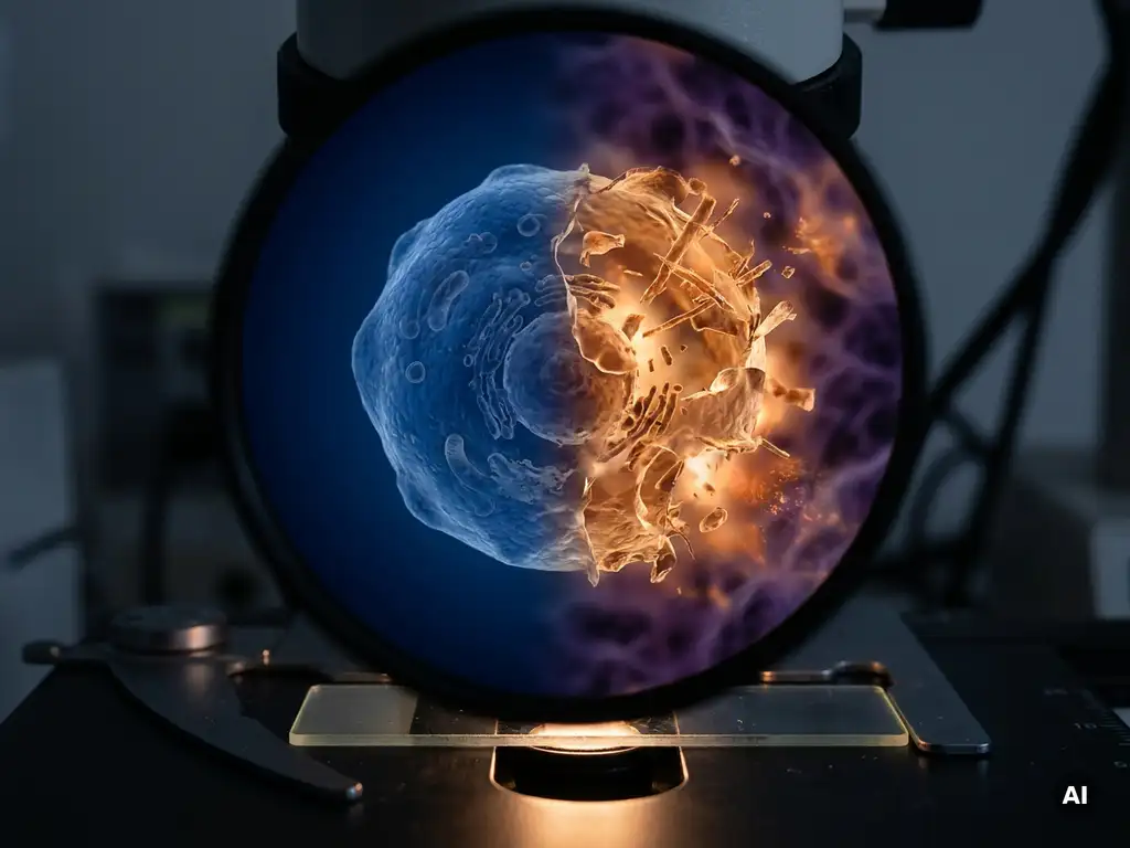
Apoptosis and necrosis are two fundamentally different forms of cell death. Apoptosis is a highly regulated, programmed process in which a cell dismantles itself in an orderly fashion without triggering inflammation. Necrosis, by contrast, is an uncontrolled form of cell death caused by injury or stress, in which the cell ruptures and releases its contents into surrounding tissue. In cancer biology, understanding the distinction between these two pathways is central to research into tumor progression and the mechanisms that cancer cells use to survive. The sections below explore each pathway in depth and explain why the difference matters for laboratory research and experimental design.
How do cancer cells resist apoptosis?
Cancer cells resist apoptosis primarily by overexpressing anti-apoptotic proteins, downregulating pro-apoptotic signals, and acquiring mutations in key regulatory genes. This resistance to programmed cell death is one of the defining hallmarks of cancer, allowing malignant cells to survive conditions that would normally trigger self-elimination.
Under normal physiological conditions, apoptosis is regulated by a balance between pro-apoptotic proteins such as BAX and BAK and anti-apoptotic proteins such as BCL-2 and BCL-XL. In many cancer types, this balance is disrupted. Elevated BCL-2 expression, for example, has been observed across a wide range of malignancies and is associated with increased cell survival.
Mutations in the TP53 gene, which encodes the tumor suppressor protein p53, are among the most frequently identified alterations in human cancers. Because p53 plays a central role in initiating apoptosis in response to DNA damage, its loss of function allows cancer cells to continue dividing even when they carry significant genomic damage.
Cancer cells also exploit survival signaling pathways, including the PI3K/AKT and RAS/ERK cascades, which promote cell proliferation and actively suppress apoptotic signals. Researchers studying these mechanisms use a range of assays, including caspase activity measurements, Annexin V staining, and mitochondrial membrane potential assays, to map how and where cancer cells interrupt the apoptotic cascade.
What triggers necrosis in cancer cells?
Necrosis in cancer cells is triggered by conditions of severe cellular stress that overwhelm the cell’s capacity to maintain homeostasis. Common triggers include hypoxia, nutrient deprivation, extreme temperature changes, physical trauma, and exposure to certain cytotoxic agents. Unlike apoptosis, necrosis is not orchestrated by the cell itself but is imposed by external conditions.
Within solid tumors, necrosis frequently occurs in the tumor core, where rapid cell proliferation outpaces the development of an adequate blood supply. The resulting oxygen and nutrient deficit leads to bioenergetic failure and cell rupture. This phenomenon is commonly observed in histological sections of aggressive tumors and is used by researchers as an indicator of tumor grade and metabolic activity.
When necrotic cells rupture, they release damage-associated molecular patterns (DAMPs) into the surrounding microenvironment. These signals can activate inflammatory responses, which in turn may influence the behavior of neighboring cancer cells and stromal tissue. Researchers studying the tumor microenvironment are increasingly interested in how necrosis-driven inflammation contributes to tumor progression and immune evasion.
It is also worth noting that a regulated form of necrotic-like cell death, termed necroptosis, has attracted growing research interest. Necroptosis shares morphological features with necrosis but is mediated by specific signaling proteins, including RIPK3 and MLKL, and represents a distinct pathway that some cancer cells may activate or suppress depending on context.
What are the key differences between apoptosis and necrosis at the cellular level?
At the cellular level, apoptosis and necrosis differ fundamentally in their morphology, biochemistry, and downstream effects on surrounding tissue. Apoptosis is characterized by cell shrinkage, chromatin condensation, membrane blebbing, and the formation of apoptotic bodies that are cleared by phagocytes without inflammation. Necrosis involves cell swelling, membrane rupture, and the release of intracellular contents that trigger local inflammation.
The following key distinctions are relevant for researchers designing cell death assays:
- Cell morphology: Apoptotic cells shrink and fragment into membrane-enclosed bodies. Necrotic cells swell and lose membrane integrity.
- Membrane integrity: Apoptosis preserves the plasma membrane until late stages. Necrosis involves early membrane disruption, making cells permeable to dyes such as propidium iodide.
- Inflammation: Apoptosis is immunologically silent. Necrosis releases pro-inflammatory signals that recruit immune cells to the site of damage.
- Energy dependence: Apoptosis is an active, ATP-dependent process. Necrosis is largely passive and occurs when ATP production fails.
- Caspase involvement: Apoptosis is driven by caspase activation. Necrosis typically proceeds without caspase activity, though necroptosis involves specific kinase signaling.
- DNA fragmentation: Apoptosis produces characteristic internucleosomal DNA laddering detectable by gel electrophoresis or TUNEL assay. Necrosis produces random, non-specific DNA degradation.
These differences have direct implications for how researchers interpret cell viability data and select appropriate detection methods for their experimental models.
Why does the type of cell death matter for cancer treatment?
The type of cell death a cancer cell undergoes has significant implications for how researchers understand tumor biology and evaluate experimental interventions in the laboratory. Whether a cell dies by apoptosis or necrosis determines the downstream immunological response, the impact on the surrounding tumor microenvironment, and the mechanistic conclusions that can be drawn from an experiment.
From a research perspective, compounds or conditions that induce apoptosis in cancer cells are generally considered more controlled and mechanistically informative than those that cause necrosis. Apoptotic death involves discrete molecular checkpoints that researchers can target and measure, making it easier to establish cause-and-effect relationships in experimental models.
Necrosis, particularly when it occurs in vivo within tumor tissue, can complicate the interpretation of experimental results. The inflammatory signals released by necrotic cells may alter the behavior of surrounding cells, introduce confounding variables in assay readouts, and affect the reproducibility of results across experimental replicates.
Researchers working in oncology-focused laboratories in the Netherlands, Belgium, and Luxembourg are increasingly investigating how the balance between apoptotic and necrotic cell death within tumor models influences the activity of the immune system. This is particularly relevant in the context of immunogenic cell death, a form of apoptosis that can stimulate anti-tumor immune responses and is an active area of preclinical investigation.
How do researchers detect and measure apoptosis versus necrosis in the lab?
Researchers detect and distinguish apoptosis from necrosis using a combination of fluorescence-based assays, flow cytometry, imaging techniques, and biochemical markers. The choice of method depends on the experimental model, the level of throughput required, and whether single-cell or population-level data are needed.
The most widely used approach for distinguishing between the two processes is the Annexin V and propidium iodide (PI) dual-staining assay, typically analyzed by flow cytometry. Annexin V binds to phosphatidylserine, which is externalized on the plasma membrane during early apoptosis, while PI only enters cells with compromised membranes. This combination allows researchers to classify cells as viable, early apoptotic, late apoptotic, or necrotic based on their staining profile.
The following numbered steps outline a general laboratory workflow for cell death discrimination:
- Harvest and wash cells to remove debris and dead cell fragments from the culture medium.
- Incubate cells with Annexin V conjugate and PI in binding buffer at room temperature, protected from light.
- Analyze immediately by flow cytometry or high-content imaging to prevent signal drift caused by ongoing cell death.
- Gate populations based on Annexin V and PI positivity to distinguish early apoptosis, late apoptosis, and necrosis.
- Validate findings with orthogonal assays such as caspase-3/7 activity measurement, TUNEL staining, or LDH release assays to confirm the cell death mechanism.
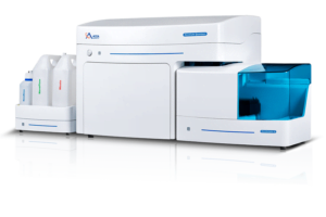 BioSPX specifically lists apoptosis assays as an application for its NovoCyte flow cytometers, as they can quantify cell-death populations and distinguish apoptosis from necrosis. As an example, the Quanteon has 4 lasers and up to 27 parameters, making it more than capable of handling typical apoptosis/necrosis flow-cytometry panels.
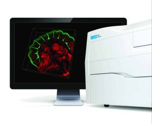
High-content imaging platforms provide spatially resolved data that can capture morphological changes associated with both apoptosis and necrosis across large cell populations, making them well suited to multi-parameter experiments. The Cytation C10 combines high-content imaging with quantitative cellular analysis, enabling researchers to visualize and characterize morphological changes associated with apoptosis and necrosis while assessing cell viability. Caspase activity assays, mitochondrial membrane potential probes, and cytochrome c release assays add mechanistic depth to cell death profiling workflows.
Can apoptosis switch to necrosis in cancer cells?
Yes, apoptosis can switch to necrosis in cancer cells under specific conditions, a phenomenon sometimes referred to as secondary necrosis or apoptosis-to-necrosis transition. This switch typically occurs when the apoptotic machinery is incomplete or overwhelmed, when phagocytic clearance of apoptotic bodies is absent, or when cellular energy levels drop too low to sustain the ATP-dependent apoptotic process.
In laboratory models, this transition is relevant for researchers who need to interpret cell death data accurately. If apoptotic cells are not efficiently cleared in culture, they may undergo secondary necrosis, releasing their contents and generating inflammatory signals that can confound experimental readouts. This is particularly important in high-density cultures or long-duration experiments where dead cell accumulation is more likely.
In cancer cells specifically, the switch between pathways may also be influenced by the activity of anti-apoptotic proteins. When caspase activation is blocked, for example by BCL-2 overexpression or caspase inhibitors, cells exposed to cytotoxic stimuli may default to a necrotic or necroptotic death rather than completing the apoptotic program. Researchers investigating this crossover use inhibitors of specific pathway components alongside dual-staining assays to map where and how the transition occurs.
Understanding the conditions under which cancer cells shift between these death pathways is an active area of research that may help investigators design more controlled and reproducible in vitro models of tumor cell death.
How BioSPX supports apoptosis and necrosis research
Accurate discrimination between apoptosis and necrosis requires instruments and workflows that deliver reliable, reproducible data across a range of experimental conditions. BioSPX supports research teams in Belgium, the Netherlands, and Luxembourg with a portfolio of laboratory instruments suited to cell death research, including:
- Multimode readers for fluorescence- and luminescence-based cell death assays, including caspase activity and LDH release measurements
- High-content imaging and microscopy systems for morphological analysis of apoptotic and necrotic cell populations
- Liquid handling solutions that support consistent, reproducible assay preparation across multiple samples
- Automation systems for cell culture maintenance, reducing variability in experimental conditions
BioSPX’s product specialists can advise on instrument selection and experimental setup to help research teams build robust cell death profiling workflows. To discuss your laboratory’s requirements, contact the BioSPX team directly.
Frequently Asked Questions
What is the difference between necroptosis and classical necrosis, and how do I know which one is occurring in my experiment?
Necroptosis is a regulated, programmed form of cell death that shares morphological features with classical necrosis — including cell swelling and membrane rupture — but is driven by specific signaling proteins, particularly RIPK3 and MLKL. To distinguish between the two, researchers typically use pharmacological inhibitors such as necrostatin-1 (a RIPK1 inhibitor) or GSK’872 (a RIPK3 inhibitor) alongside standard necrosis markers like LDH release and PI uptake. If inhibiting these kinases reduces cell death, necroptosis is likely involved rather than passive necrosis.
Which cell death assay should I prioritize if I'm working with a limited sample size or low cell numbers?
For low cell number samples, caspase-3/7 luminescence-based assays are often the most sensitive option, as they require fewer cells than flow cytometry-based Annexin V/PI protocols and can be run in a multiwell plate format on a standard multimode reader. If morphological data is also needed, high-content imaging allows you to extract both quantitative and spatial information from the same small sample. Combining a luminescent caspase assay with an LDH release measurement in parallel wells is a practical, resource-efficient approach for confirming both apoptotic and necrotic contributions.
How can I minimize secondary necrosis artifacts when running long-duration or high-density cell death experiments?
To reduce secondary necrosis in culture, collect and analyze samples at regular, shorter time intervals rather than at a single late endpoint, and avoid allowing dead cells to accumulate in the culture medium. Centrifuging and resuspending cells in fresh binding buffer just before staining helps remove debris that can interfere with Annexin V/PI gating. In high-density cultures, consider switching to a high-content imaging platform, which allows you to spatially resolve and exclude debris from your analysis rather than relying solely on population-level flow cytometry data.
Are there cancer cell lines that are particularly well suited for studying apoptosis resistance mechanisms?
Several well-characterized cell lines are commonly used as models of apoptosis resistance, including BCL-2-overexpressing lines such as certain B-cell lymphoma models, and TP53-mutant lines such as MDA-MB-231 (breast) or DU145 (prostate). These lines allow researchers to study specific resistance mechanisms in isolation and are widely referenced in the literature, making it easier to benchmark results. When selecting a model, it is important to verify the expression status of key apoptotic regulators — BCL-2, BAX, p53, and caspase-8 — in your specific cell line passage, as these can drift over time.
Can the tumor microenvironment influence whether cancer cells die by apoptosis or necrosis in my in vitro model?
Yes, microenvironmental factors such as oxygen tension, glucose availability, and pH can significantly shift the balance between apoptotic and necrotic cell death, even in standard cell culture models. Hypoxic conditions, for example, can deplete ATP to levels that prevent the completion of the energy-dependent apoptotic program, pushing cells toward necrosis. Researchers aiming to model tumor core conditions more accurately can use hypoxia chambers or 3D spheroid culture systems, which more faithfully replicate the metabolic gradients found in solid tumors and produce more physiologically relevant cell death readouts.
What controls should I always include when running an apoptosis versus necrosis discrimination assay?
Every cell death discrimination experiment should include at minimum four controls: an untreated viable cell population, a positive apoptosis control (such as staurosporine-treated cells), a positive necrosis control (such as heat-killed or detergent-lysed cells), and a single-stain compensation control for each fluorophore used in the assay. These controls allow you to correctly set gates in flow cytometry analysis and validate that your Annexin V and PI reagents are performing as expected. Including a caspase inhibitor control (e.g., Z-VAD-FMK) is also recommended if you want to confirm that the apoptotic signal is caspase-dependent.
How does immunogenic cell death differ from standard apoptosis, and why is it relevant to current oncology research?
Immunogenic cell death (ICD) is a specialized form of apoptosis in which dying cancer cells emit specific danger signals — including calreticulin exposure on the cell surface, ATP secretion, and HMGB1 release — that actively stimulate an anti-tumor immune response. Unlike conventional apoptosis, which is immunologically silent, ICD can prime dendritic cells and activate cytotoxic T lymphocytes against tumor antigens. This makes ICD particularly relevant in the context of cancer immunotherapy research, where preclinical models are being used to identify compounds or treatment combinations that can convert immunologically ‘cold’ tumors into ones capable of driving adaptive immune responses.
Related Articles
- How can researchers quantify FRET across heterogeneous cell populations?
- How does angiogenesis support tumor growth at the cellular level?
- How do oncogenes differ from tumor suppressor genes in cancer development?
- What causes cancer cells to behave differently across repeated experiments?
- How does cancer spread to other parts of the body?
This content was generated with the help of AI and it may contain mistakes