
 EN
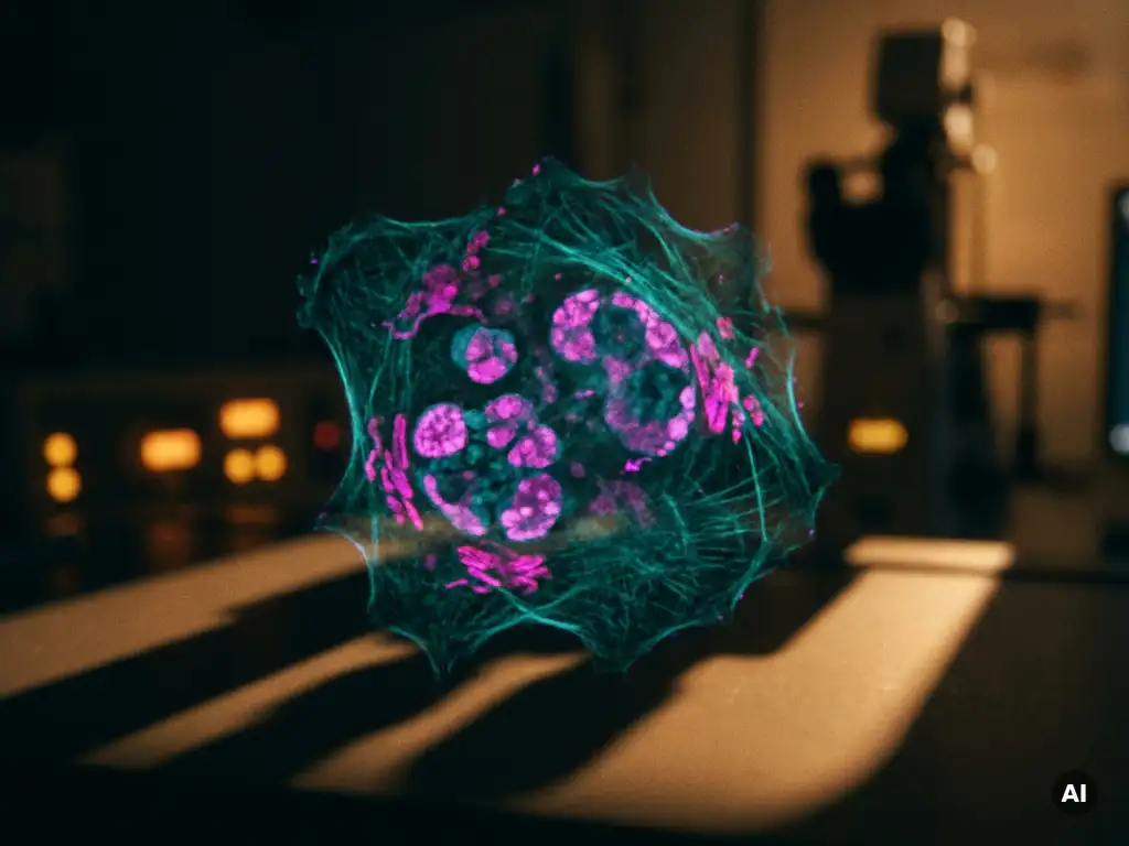
Synthetic lethality is a genetic phenomenon in which the simultaneous loss of function of two genes causes cell death, while the loss of either gene alone is tolerated. In cancer research, this principle is exploited to selectively kill tumour cells that already carry a specific genetic defect, leaving healthy cells largely unaffected. The sections below unpack the key questions researchers are asking about this approach in 2026.
How does synthetic lethality kill cancer cells?
Synthetic lethality kills cancer cells by targeting a second gene or pathway that the cell depends on for survival, given that a first gene is already non-functional due to a cancer-driving mutation. Because healthy cells retain a functional copy of the first gene, they can tolerate the loss of the second. Tumour cells, lacking that backup, cannot survive and undergo cell death.
This selectivity is what makes synthetic lethality such a compelling framework in oncology research. Rather than broadly attacking dividing cells, as conventional chemotherapy does, the synthetic lethal approach is designed around the specific genetic vulnerabilities that define a tumour. Researchers can, in principle, identify which gene pairs produce a synthetic lethal interaction and then develop inhibitors that target the surviving partner gene in patients whose tumours already carry the first mutation.
The concept was first described in model organisms such as yeast and fruit flies, where systematic genetic screens revealed thousands of gene pairs whose combined loss was lethal. Advances in CRISPR-based screening and high-throughput genomics have since made it possible to map these interactions in human cancer cell lines at scale.
What genes and pathways are most commonly targeted?
The most extensively studied synthetic lethal interactions in cancer involve genes in the DNA damage response (DDR) network. Mutations in BRCA1 and BRCA2 are the best-characterised examples, as these genes are essential for high-fidelity repair of double-strand DNA breaks through homologous recombination. Tumours harbouring BRCA mutations are therefore dependent on alternative repair mechanisms for survival.
Beyond BRCA, researchers are actively investigating synthetic lethal relationships involving:
- ATM, ATR, and CHK1/CHK2 – kinases that coordinate the DNA damage checkpoint response
- RAD51 and PALB2 – proteins involved in homologous recombination repair
- RB1 and TP53 – tumour suppressor genes whose loss creates dependencies in cell cycle regulation
- KRAS and STK11 – oncogene and tumour suppressor combinations relevant to lung and pancreatic cancers
- WRN helicase – recently identified as a promising target in microsatellite-unstable tumours
The DDR pathway remains central because DNA repair is a fundamental survival mechanism, and many tumours accumulate mutations in these genes during progression. Researchers in the Benelux and globally are expanding the search to metabolic pathways, chromatin-remodelling complexes, and RNA processing machinery, broadening the landscape of potential cancer drug targets.
What are PARP inhibitors and how do they exploit synthetic lethality?
PARP inhibitors are small molecules that block poly(ADP-ribose) polymerase enzymes, which play a key role in repairing single-strand DNA breaks. In tumour cells with BRCA1 or BRCA2 mutations, inhibiting PARP creates a synthetic lethal situation: unrepaired single-strand breaks collapse into double-strand breaks, and without functional homologous recombination, the cancer cell cannot repair them and dies.
PARP inhibitors represent the most clinically advanced application of synthetic lethality and have become an important tool for studying DDR biology in research settings. From a laboratory perspective, they serve as both research probes to dissect repair pathway dependencies and reference compounds in drug discovery screens.
Researchers use PARP inhibitors experimentally to:
- Validate homologous recombination deficiency in cell line models
- Investigate mechanisms of resistance that emerge after prolonged exposure
- Explore combination strategies with other DDR inhibitors or immunological approaches
- Develop and refine biomarker assays that predict sensitivity to this class of compounds
Understanding resistance mechanisms is a particularly active area of investigation, as tumour cells can restore homologous recombination capacity through secondary mutations or epigenetic reprogramming, which reduces the effectiveness of PARP inhibition over time. Studying resistance means following cells over weeks, not days. Automated culture systems such as Cellaven’s Nestor can handle repeated feeding and passaging without weekend shifts, while in-incubator live-cell imagers (Curiosis Celloger) and long-term metabolism monitoring (Leadgene DolphinQ) show how cells adapt under sustained inhibitor exposure. To see whether repair complexes still interact in situ, HAWK’s QF-Pro® reagent kits make protein–protein interactions and post-translational modifications quantifiable in cells and tissue.
How do researchers identify new synthetic lethal interactions?
Researchers identify new synthetic lethal interactions primarily through large-scale genetic screens, most commonly using CRISPR-Cas9 or RNA interference libraries to systematically silence genes across cancer cell lines and measure which combinations reduce viability. Computational analysis of cancer genomics datasets can then prioritise interactions that are statistically enriched in tumours carrying specific mutations.
High-throughput functional genomics has transformed this field. Projects such as large-scale cancer dependency mapping efforts have profiled hundreds of cancer cell lines to generate comprehensive maps of genetic vulnerabilities. These datasets are increasingly used by research groups in Belgium, the Netherlands, and Luxembourg to guide hypothesis generation and experimental design.
Complementary approaches include:
- Compound library screens combined with isogenic cell line pairs that differ only in the gene of interest
- Proteomics and transcriptomics to identify compensatory pathway activation after gene loss
- Organoid models derived from patient tumour tissue, which preserve more of the genetic complexity of real tumours than standard cell lines
- Machine learning models trained on existing interaction data to predict untested gene pairs
Each approach has strengths and limitations. Cell line screens are scalable but may not capture the tumour microenvironment or stromal interactions. Once candidate gene pairs emerge from a screen, they need orthogonal validation. Label-free impedance monitoring, such as the xCELLigence RTCA platform, tracks proliferation, morphology and attachment continuously, so researchers can see when a synthetic lethal effect starts rather than relying on a single endpoint. Flow cytometry (for example the NovoCyte series) adds multiparametric readouts of apoptosis, cell-cycle arrest and DNA-damage markers, while imaging readers such as the BioTek Cytation 9 or C10 capture phenotypic changes under controlled temperature and gas conditions. For larger compound or guide panels, automated reagent dispensing (BioTek MultiFlo FX) and automated incubation (BioSpa 8) help keep plate-to-plate variability low.
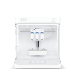
Organoid-based models offer greater biological relevance but are more resource-intensive and harder to run at the throughput needed for genome-wide discovery. Practical bottlenecks, such as the labour and cost of maintaining organoids, are being addressed by dedicated hardware. The Orgonex RPMotion bioreactor, for instance, is a compact system designed for organoid expansion, with the manufacturer reporting faster growth and reduced hands-on time. Bioprinting platforms such as CELLINK’s Bio X series offer a complementary route to building reproducible 3D tumour models, and these are best seen as an addition to existing model systems rather than a replacement.
What are the main challenges in translating synthetic lethality to the clinic?
The main challenges in translating synthetic lethality from the laboratory to clinical research include tumour heterogeneity, the development of resistance mechanisms, the difficulty of identifying reliable predictive biomarkers, and the complexity of validating candidate interactions in models that accurately reflect human tumour biology.
Tumour heterogeneity is a fundamental obstacle. Even within a single tumour, different cell populations may carry distinct mutation profiles, meaning that a synthetic lethal interaction present in one subclone may be absent in another. This can allow resistant populations to expand under selective pressure.
Biomarker development is equally challenging. Because bulk measurements can mask resistant subclones, single-cell readouts are increasingly valuable. Single-cell analysis and high-parameter flow cytometry can reveal subpopulations that respond differently to the same perturbation, while in situ interaction assays help link these differences to tissue context. For a synthetic lethal strategy to be applied in research or drug development, researchers need robust assays that can accurately classify tumours as deficient in the relevant pathway. Homologous recombination deficiency scoring, for example, remains an active area of assay development, with ongoing work to standardise methods and improve sensitivity across different tissue types.
Additional challenges include the fact that many synthetic lethal interactions identified in cell lines do not replicate in more complex models, and that the genetic background of a tumour can modulate the strength of a given interaction in ways that are difficult to predict.
Which cancer types benefit most from synthetic lethality approaches?
Cancer types with high rates of DNA damage response gene mutations are currently the most studied in the context of synthetic lethality. These include ovarian, breast, pancreatic, and prostate cancers, where BRCA1 and BRCA2 mutations are relatively common. Colorectal and endometrial cancers with microsatellite instability are also of growing research interest due to their distinct repair deficiencies.
Researchers are working to extend the synthetic lethal framework beyond BRCA-mutated tumours. Lung cancer, particularly subtypes driven by specific oncogene mutations, and haematological malignancies with defined genetic lesions are active areas of investigation. The broader goal is to identify actionable synthetic lethal pairs across a much wider range of tumour types, which would expand the research and therapeutic utility of this approach considerably.
For research groups in the Benelux working on cancer biology, the diversity of tumour types under investigation means that experimental platforms need to be flexible. Models ranging from two-dimensional cell cultures to three-dimensional organoid systems and co-culture formats are all in use, depending on the biological question being addressed. Whichever model is chosen, the readout platform needs to follow it. Imaging readers with environmental control support live co-culture experiments, while the same plate-based workflows can often be carried from 2D into 3D formats.
How BioSPX supports synthetic lethality and oncology research
Investigating synthetic lethality requires precise, reproducible experimental platforms capable of handling complex cell models and high-throughput screening workflows. BioSPX supports research groups in Belgium, the Netherlands, and Luxembourg with instruments and expertise suited to these demands.
- Multimode readers and imaging systems for viability, apoptosis, and DNA damage assays across well plate formats
- Liquid handling solutions to support consistent, reproducible compound addition in screening workflows
- Organoid and 3D culture tools, including bioreactor systems for more physiologically relevant tumour models
- Automated cell culture platforms to reduce manual variability in long-term experiments
- Expert product specialists who advise on instrument selection and experimental configuration
- Installation, training, and maintenance support from a dedicated service team
If your research group is building or scaling a synthetic lethality programme, contact BioSPX to discuss which instruments and workflows best fit your experimental needs.
Frequently Asked Questions
How do I choose the right cell line model for studying a specific synthetic lethal interaction?
The choice of cell line model depends on the genetic mutation you want to study and the biological question you are asking. Isogenic cell line pairs — where two lines are identical except for the gene of interest — are the gold standard for isolating a specific synthetic lethal interaction, as they minimise confounding genetic background effects. For interactions involving complex tumour microenvironments or stromal signalling, patient-derived organoid models offer greater biological relevance, though they require more resource-intensive workflows. Consulting cancer dependency mapping datasets can also help you identify well-characterised cell lines that are already confirmed to carry the mutation profile relevant to your research.
What is 'BRCA-ness' or homologous recombination deficiency (HRD), and why does it matter beyond BRCA1/2 mutations?
Homologous recombination deficiency (HRD), sometimes called ‘BRCAness,’ refers to a functional state in which a tumour cell cannot efficiently perform homologous recombination repair, regardless of whether BRCA1 or BRCA2 are directly mutated. Mutations in other HR pathway genes such as PALB2, RAD51, or BRIP1 can produce the same functional deficiency and, in principle, the same synthetic lethal vulnerability to PARP inhibition. This distinction matters because it significantly expands the patient population that may benefit from DDR-targeting strategies, and it underscores the importance of functional HRD assays over simple mutation-detection approaches when classifying tumours for research or drug development purposes.
What are the most common reasons a synthetic lethal interaction identified in a screen fails to validate in follow-up experiments?
The most frequent reasons for failed validation include genetic background variability between cell lines, off-target effects from the RNAi or CRISPR reagents used in the primary screen, and differences in assay conditions such as drug concentration, treatment duration, or growth format. A synthetic lethal interaction can also appear in a screen but be too weak or context-dependent to reproduce consistently — for example, it may only manifest under specific metabolic or microenvironmental conditions not replicated in standard culture. Using multiple independent gene-silencing approaches, orthogonal assay readouts, and at least one isogenic cell line pair during validation significantly improves confidence in a candidate interaction before committing to deeper mechanistic studies.
How can machine learning and computational tools realistically help a wet-lab research group working on synthetic lethality?
For wet-lab groups, the most practical entry point into computational tools is using publicly available cancer dependency datasets — such as those from large-scale dependency mapping projects — to prioritise which gene pairs are worth testing experimentally, rather than attempting genome-wide screens from scratch. Several user-friendly web portals allow researchers to query gene dependencies across hundreds of cell lines without requiring bioinformatics expertise. More advanced machine learning models trained on existing synthetic lethal interaction data can predict novel pairs, but these outputs are best treated as hypothesis-generators that still require rigorous experimental validation, particularly in models that reflect the genetic context of the tumour type you are studying.
What assays are most useful for confirming that a compound is inducing synthetic lethality rather than general cytotoxicity?
The key to distinguishing synthetic lethality from general cytotoxicity is demonstrating selectivity — the compound should reduce viability specifically in cells carrying the relevant mutation, while cells with an intact copy of that gene remain largely unaffected. A standard approach is to compare dose-response curves in isogenic cell line pairs (mutant vs. wild-type) using viability assays such as CellTiter-Glo or resazurin-based methods. Complementary assays measuring apoptosis markers (e.g., caspase activation or Annexin V staining), DNA damage markers such as γH2AX foci, and colony formation assays help confirm that the observed cell death is mechanistically consistent with the expected synthetic lethal pathway rather than off-target toxicity.
Are there any practical strategies for studying resistance mechanisms to synthetic lethal interactions in the lab?
The most widely used approach is generating drug-resistant cell lines through prolonged exposure to a synthetic lethal compound — such as a PARP inhibitor — at sub-lethal concentrations, then characterising the resistant clones using whole-exome sequencing, transcriptomics, or proteomics to identify the resistance mechanism. Comparing the molecular profiles of resistant and sensitive clones can reveal secondary mutations that restore the originally deficient pathway, or compensatory pathway activations that bypass the synthetic lethal dependency. CRISPR-based suppressor screens, where a genome-wide library is introduced into already-treated cells to identify genes whose loss restores viability, are also a powerful and increasingly accessible strategy for systematically mapping resistance mechanisms.
How should research groups in the Benelux think about scaling up from initial hit identification to a more robust synthetic lethality programme?
Scaling up typically requires moving from manual, low-throughput validation experiments toward more automated and reproducible workflows, particularly for compound screening, dose-response profiling, and multi-parameter imaging assays. Investing in liquid handling automation early reduces pipetting variability — a common source of noise in viability screens — and enables consistent compound addition across larger plate formats. It is also worth establishing a diverse panel of cell models at the outset, including at least one organoid system relevant to your tumour type of interest, so that promising hits can be tested in progressively more complex biological contexts before significant resources are committed to any single candidate interaction. Partnering with instrument specialists familiar with oncology screening workflows can help research groups configure platforms that are both scalable and suited to their specific experimental needs.
Related Articles
- How does FRET measure molecular proximity below the resolution limit of conventional microscopy?
- Why is fluorescence lifetime imaging useful for studying protein interactions?
- What is the tumor microenvironment and why does it matter in research?
- What causes cancer cells to behave differently across repeated experiments?
- What is the difference between 2D and 3D tumor cell culture models?
This content was generated with the help of AI and it may contain mistakes