
 EN
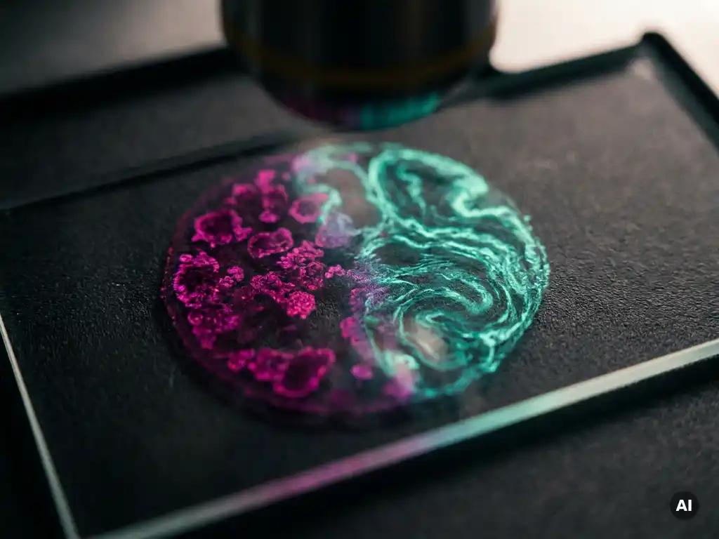
Spatial proteomics and spatial transcriptomics are complementary but distinct approaches to mapping biological information within tissue. Spatial proteomics measures the location and abundance of proteins directly, while spatial transcriptomics captures the spatial distribution of messenger RNA transcripts, reflecting gene expression activity. The two methods target different molecular layers of the same biological system, which means each answers a different set of research questions. The sections below explore how they differ technically, where each excels, and when combining them delivers the most complete picture of tissue biology.
How do spatial proteomics and spatial transcriptomics capture biological information differently?
Spatial proteomics captures protein identity, abundance, and localization directly within tissue sections, while spatial transcriptomics measures RNA transcript levels at defined spatial coordinates. The core distinction is the molecular target: proteins are the functional effectors of biology, whereas transcripts represent the intermediate step between gene activation and protein production. This means the two approaches interrogate the same tissue at different molecular levels.
In spatial proteomics workflows, antibody-based detection methods such as multiplexed immunofluorescence or mass spectrometry imaging are used to identify and quantify proteins while preserving their spatial context within the tissue architecture. The result is a map of where specific proteins are expressed, co-localized, or depleted across cell populations and microenvironments.
Spatial transcriptomics, by contrast, typically relies on sequencing-based or hybridization-based methods to detect RNA transcripts at defined positions within a tissue section. Approaches range from in situ hybridization to barcoded capture platforms that assign transcript reads to spatial coordinates. The output is a gene expression profile anchored to tissue location, revealing which genes are active in which regions.
Because transcript levels do not always correlate directly with protein abundance due to post-transcriptional regulation, protein turnover, and translational efficiency, the two methods can yield meaningfully different biological pictures of the same sample.
What biological questions can only spatial proteomics answer?
Spatial proteomics is the only approach that can directly measure protein-level events within tissue, including post-translational modifications, protein-protein interactions, and the activation states of signaling pathways. These molecular events are invisible to transcriptomic methods because they occur after translation and leave no RNA signature.
Researchers use spatial proteomics to investigate questions such as:
- Where within a tumor microenvironment are specific immune checkpoint proteins expressed, and do they co-localize with immune cell populations?
- How are phosphorylation or glycosylation patterns distributed across tissue regions, and what does this reveal about active signaling cascades?
- Which cell types are defined by their protein surface markers rather than their transcriptional state, and how are they spatially organized?
- How does protein secretion vary across tissue compartments, particularly in glandular or stromal structures?
Because many biological processes are regulated at the protein level rather than the transcript level, spatial proteomics is essential for understanding the functional state of cells and tissues rather than just their transcriptional activity.
What biological questions can only spatial transcriptomics answer?
Spatial transcriptomics is uniquely suited to capturing genome-wide gene expression patterns across tissue in an unbiased way. Unlike spatial proteomics, which is constrained by the availability of validated antibodies or mass spectrometry reagents, spatial transcriptomics can in principle survey the entire transcriptome without prior knowledge of which genes to target.
This makes spatial transcriptomics particularly powerful for questions such as:
- Which gene regulatory programs are active in specific tissue zones, and how do these programs shift across developmental stages?
- How do rare or previously uncharacterized cell populations define themselves transcriptionally, and where are they located within tissue?
- What are the spatially resolved gene expression gradients across tissue boundaries, for example at the interface between healthy and diseased regions?
- Which transcription factor networks are active in spatially defined niches, and how do they relate to cell identity?
Spatial transcriptomics also integrates naturally with single-cell RNA sequencing datasets, enabling researchers to deconvolve bulk spatial signals into inferred cell type compositions. This kind of multi-scale analysis is not easily replicated using protein-based methods alone.
How do the technical workflows and instruments compare between the two approaches?
The workflows for spatial proteomics and spatial transcriptomics differ substantially in their reagents, instrumentation, and data processing requirements. Spatial proteomics typically requires high-quality, validated antibodies and relies on imaging platforms capable of resolving multiple protein signals simultaneously within tissue sections. Spatial transcriptomics workflows center on nucleic acid chemistry, requiring tissue permeabilization, RNA capture or in situ hybridization, and sequencing or imaging readout.
Spatial proteomics instrumentation
Multiplexed protein imaging platforms use fluorescence-based or metal-tagged antibody panels to detect dozens of proteins in a single tissue section. High-content imaging systems with automated image acquisition and analysis pipelines are central to these workflows. Mass spectrometry imaging instruments offer label-free protein detection with high molecular specificity but require specialized sample preparation and data analysis expertise. The imaging and cellular analysis instruments used in these workflows must deliver high spatial resolution to distinguish signals at the subcellular level.
Spatial transcriptomics instrumentation
Spatial transcriptomics platforms range from barcoded slide-based capture systems to single-molecule fluorescence in situ hybridization approaches. Barcoded capture methods require next-generation sequencing as a readout, while imaging-based methods use fluorescence microscopes capable of detecting individual RNA molecules. Computational infrastructure for sequence alignment, spatial mapping, and downstream statistical analysis is a significant component of the workflow, often requiring bioinformatics expertise that complements the laboratory instrumentation.
When should researchers combine spatial proteomics and spatial transcriptomics?
Researchers should consider combining spatial proteomics and spatial transcriptomics when a single molecular layer is insufficient to answer the experimental question. Integrating both approaches, often called spatial multi-omics, allows researchers to examine the relationship between gene expression and protein output in the same tissue, revealing where and why discordance between transcript and protein levels occurs.
Combining the two methods is particularly valuable in the following scenarios:
- Tumor microenvironment research: Understanding how immune cell gene expression programs translate into protein-level functional states, such as cytokine secretion or receptor expression, requires both layers of information.
- Developmental biology: Mapping how transcriptional gradients during organogenesis give rise to spatially distinct protein expression domains helps researchers connect gene regulation to tissue patterning.
- Disease mechanism investigation: When transcript and protein abundance diverge in diseased tissue, multi-omics integration may reveal post-transcriptional regulatory mechanisms that drive pathology.
- Cell type validation: Transcriptomically defined cell clusters can be validated and further characterized by confirming protein marker co-expression at the same spatial locations.
While combined workflows add technical complexity and cost, the depth of insight they generate is increasingly justified as spatial biology matures as a field.
What are the main limitations of each spatial omics technology?
Both spatial proteomics and spatial transcriptomics carry significant technical limitations that researchers must account for in experimental design and data interpretation.
Spatial proteomics limitations include dependence on antibody availability and specificity, which constrains the number of proteins that can be reliably detected. Multiplexing depth is currently more limited than transcriptomic approaches, and signal overlap between fluorescent channels can reduce quantitative accuracy. Post-translational modifications are detectable only when modification-specific antibodies exist and have been validated for the tissue type in question.
Spatial transcriptomics limitations center on RNA sensitivity and spatial resolution. Barcoded capture methods may not detect low-abundance transcripts reliably, and the spatial resolution of some platforms is insufficient to assign transcripts to individual cells without computational deconvolution. RNA integrity is highly sensitive to tissue handling and fixation conditions, meaning sample quality has a major impact on data reliability. In addition, transcript abundance does not directly reflect protein levels, which limits the functional conclusions that can be drawn from transcriptomic data alone.
Both approaches generate large, complex datasets that require substantial computational resources and bioinformatics expertise to process and interpret meaningfully. Reproducibility across samples, tissue types, and laboratory settings remains an active area of methodological development across the spatial omics field.
How BioSPX supports spatial biology research in the Benelux
For research teams in Belgium, the Netherlands, and Luxembourg working with spatial proteomics or spatial transcriptomics, selecting and implementing the right instrumentation is a critical step. BioSPX supports Benelux researchers across these workflows by providing:
- Access to an extensive portfolio of imaging and microscopy systems suited to multiplexed spatial analysis
- Liquid handling and automation solutions that improve consistency in sample preparation workflows
- Expert guidance from product specialists who advise on instrument selection, experimental configuration, and workflow optimization
- Installation, user training, and responsive maintenance support to ensure instruments perform reliably over time
Whether your laboratory is establishing a spatial omics workflow for the first time or scaling an existing approach, BioSPX can help you identify the right instrumentation for your research objectives. Contact the BioSPX team to discuss your specific requirements and find the solution that best fits your work.
Frequently Asked Questions
How do I decide which spatial omics approach to start with if I have no prior experience with either?
Start by defining the specific biological question you need to answer: if you are investigating protein-level functional states, signaling activity, or post-translational modifications, spatial proteomics is the more direct choice; if you want unbiased, genome-wide gene expression mapping or need to discover novel cell populations, spatial transcriptomics is the better entry point. Consider also your existing laboratory infrastructure — if your team already has fluorescence imaging capabilities and antibody-based assay experience, spatial proteomics may have a lower barrier to entry, whereas spatial transcriptomics requires sequencing access and bioinformatics support. Starting with a single, well-defined experimental question and the technology best suited to answer it will give you more interpretable results than attempting a multi-omics approach from the outset.
What sample preparation mistakes most commonly compromise spatial omics data quality?
Inconsistent or suboptimal tissue fixation is one of the most frequent sources of data quality issues across both approaches — over-fixation with formalin can cross-link RNA and reduce transcript detection in spatial transcriptomics, while under-fixation can compromise tissue morphology and antibody binding in spatial proteomics. For spatial transcriptomics specifically, delays between tissue collection and fixation lead to RNA degradation that significantly reduces sensitivity and reproducibility. In spatial proteomics, insufficient antibody validation for the specific tissue type and fixation condition used is a common oversight that introduces false-positive or false-negative signals. Establishing a standardized tissue handling and fixation protocol before beginning a study, and validating it with control tissues, is one of the highest-impact steps a team can take.
Can spatial proteomics and spatial transcriptomics be performed on the same tissue section?
Performing both assays on the exact same tissue section is technically challenging but increasingly feasible with emerging co-detection protocols and sequential staining workflows. More commonly, researchers perform the two assays on serial sections cut from the same tissue block, then computationally co-register the resulting spatial maps to align protein and transcript data to a shared coordinate system. True same-section multi-omics approaches exist — some platforms are being developed specifically to enable simultaneous or sequential protein and RNA detection — but they currently involve significant protocol optimization and may involve trade-offs in sensitivity for one or both molecular layers. For most laboratories, serial section co-registration remains the most practical and reproducible strategy.
How many protein targets can spatial proteomics realistically detect in a single experiment, and how does that compare to spatial transcriptomics?
Current multiplexed immunofluorescence platforms can typically detect between 20 and 60 protein targets per tissue section in a single experiment, while mass spectrometry imaging approaches can extend this to hundreds of proteins but with different spatial resolution and throughput trade-offs. Spatial transcriptomics, particularly sequencing-based barcoded capture methods, can in principle capture tens of thousands of transcripts simultaneously, giving it a substantial advantage in molecular breadth. Imaging-based spatial transcriptomics panels are more targeted but can still profile hundreds to thousands of genes at once. This difference in multiplexing depth is one of the key practical considerations when choosing between the two approaches, especially for discovery-oriented versus hypothesis-driven research.
How should I handle the bioinformatics and data analysis requirements if my lab lacks dedicated computational expertise?
Both spatial proteomics and spatial transcriptomics generate large, complex datasets, but several well-documented open-source tools — including Seurat, Squidpy, and FIJI-based image analysis pipelines — provide accessible starting points for researchers without deep bioinformatics backgrounds. Many instrument vendors also provide proprietary analysis software with graphical interfaces that lower the computational barrier for standard analyses such as cell segmentation, clustering, and spatial neighborhood analysis. Collaborating with a bioinformatics core facility or a computational biology colleague is strongly advisable for more advanced analyses such as multi-omics data integration or spatial statistical modeling. Investing time in understanding the data structure your platform produces before beginning a large study will make downstream analysis significantly more manageable.
Is spatial transcriptomics data directly comparable across different platforms or studies?
Cross-platform and cross-study comparability in spatial transcriptomics is an active challenge in the field, as differences in spatial resolution, capture efficiency, tissue handling protocols, and sequencing depth can all introduce technical variation between datasets. Normalization strategies and batch correction methods adapted from single-cell RNA sequencing are commonly applied, but their effectiveness depends heavily on study design and the degree of technical divergence between datasets. When designing a study intended for comparison with published data, carefully matching tissue fixation protocols, section thickness, and analysis pipelines to those used in the reference dataset will improve comparability. Reporting full methodological details in publications is also critical for enabling meaningful cross-study interpretation.
What are the most important questions to ask a vendor or instrument specialist before investing in a spatial omics platform?
Key questions to ask include: what is the validated spatial resolution of the platform and is it sufficient to resolve individual cells or subcellular compartments in your tissue type? What is the realistic multiplexing depth for your target molecule class, and what reagents or panels are already validated for your specific tissue and fixation conditions? Ask also about the full workflow — including sample preparation requirements, data acquisition time per section, and the computational infrastructure needed for analysis — to assess total resource requirements beyond the instrument itself. Finally, ask about the vendor’s support model, including training, troubleshooting access, and software update timelines, as ongoing technical support is a significant factor in long-term workflow success.
Related Articles
- How can researchers quantify FRET across heterogeneous cell populations?
- How can protein interactions be mapped across different subcellular compartments?
- What is label-free cell analysis and how does it work in oncology research?
- What is the role of high-content imaging in cancer cell phenotyping?
- What is the difference between 2D and 3D tumor cell culture models?
This content was generated with the help of AI and it may contain mistakes