
 EN
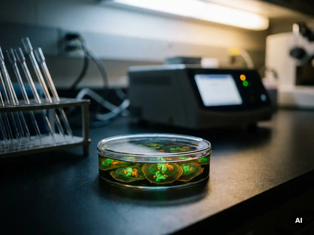
Protein interactions can be mapped across different subcellular compartments by combining compartment-specific isolation techniques with interaction detection methods such as proximity labeling, co-immunoprecipitation, FRET, BRET, and organelle proteomics. Because the cellular environment varies dramatically between the nucleus, mitochondria, endoplasmic reticulum, and other organelles, no single method captures all interaction contexts equally well. The sections below address the key approaches, their underlying principles, and the instrumentation that supports this research.
Which methods reveal protein interactions in specific organelles?
Several complementary methods can reveal protein-protein interactions within specific organelles, including co-immunoprecipitation (co-IP) following organelle fractionation, proximity labeling (such as BioID and APEX), split-complementation assays, and fluorescence-based energy transfer techniques. Each method offers a different balance between spatial resolution, sensitivity, and compatibility with live or fixed cell preparations.
Co-immunoprecipitation is one of the most widely used approaches for detecting stable protein complexes. When combined with subcellular fractionation, it allows researchers to enrich a specific organelle fraction before pulling down the protein of interest, which reduces background from non-relevant compartments. However, co-IP captures primarily stable interactions and may miss transient or low-affinity associations.
Split-complementation assays, such as split-GFP or split-luciferase systems, offer an alternative strategy. In these assays, two non-functional fragments of a reporter protein are each fused to a candidate interaction partner. When the two proteins come into close proximity within a specific compartment, the fragments reconstitute a functional signal. These assays can be targeted to specific organelles by attaching appropriate localization sequences, making them particularly useful for studying interactions in compartments that are difficult to isolate biochemically.
How does proximity labeling map compartment-specific interactions?
Proximity labeling maps compartment-specific protein interactions by fusing an enzymatic label, such as a biotin ligase (BioID, TurboID) or a peroxidase (APEX2), to a protein of interest or an organelle-targeting sequence. The enzyme biotinylates all proteins within a defined radius, typically a few nanometers, which can then be captured by streptavidin pulldown and identified by mass spectrometry.
The key advantage of proximity labeling is that it captures transient and weak interactions that co-IP often misses. Because labeling occurs inside the living cell, the interaction landscape reflects the native cellular environment rather than conditions introduced during cell lysis. Researchers can direct the labeling enzyme to a specific compartment, for example, the mitochondrial matrix or the inner nuclear membrane, by fusing it to a well-characterized targeting signal.
The resulting datasets provide a spatial map of the local proteome surrounding the bait protein within that compartment. When combined with quantitative mass spectrometry, proximity labeling can distinguish proteins that are consistently present near the bait from those that appear only under specific experimental conditions, such as after a signaling stimulus or stress response. This makes proximity labeling a particularly powerful tool for studying how subcellular interaction networks reorganize dynamically.
What is the difference between FRET and BRET for live-cell interaction studies?
FRET (Förster Resonance Energy Transfer) and BRET (Bioluminescence Resonance Energy Transfer) both detect protein-protein interactions by measuring energy transfer between a donor and an acceptor fluorophore when two proteins are within approximately 10 nanometers of each other. The key difference is the energy source: FRET uses an external excitation light source, while BRET uses a bioluminescent enzyme as the donor, eliminating the need for external illumination.
In FRET experiments, the donor fluorophore is excited by a laser or lamp, and if the acceptor is sufficiently close, energy is transferred non-radiatively, producing a detectable acceptor emission signal. FRET is well-suited to high-resolution imaging because the excitation light can be focused to specific subcellular regions, allowing researchers to visualize interactions at defined locations within a cell.
BRET avoids the phototoxicity and autofluorescence that can complicate FRET measurements in live cells. Because the donor signal is generated enzymatically, BRET is particularly useful for studying interactions in compartments where background fluorescence is high or where prolonged imaging would damage the cells. BRET is also more amenable to plate-based, higher-throughput formats, making it suitable for screening interaction conditions across many samples.
Both approaches can be targeted to specific subcellular compartments by attaching appropriate localization sequences to the fusion proteins, enabling researchers to confirm not only that an interaction occurs but also where within the cell it takes place.
How can co-fractionation and organelle proteomics identify compartment interactions?
Co-fractionation proteomics identifies compartment-specific protein interactions by separating cellular contents into fractions according to density, size, or biochemical properties, then using mass spectrometry to determine which proteins consistently co-migrate across multiple fractions. Proteins that share a fractionation profile are likely to be part of the same complex or to reside in the same organelle.
Organelle proteomics extends this principle by using techniques such as density gradient centrifugation, free-flow electrophoresis, or immunoisolation to enrich specific organelles before proteomic analysis. Each enrichment step improves the signal-to-noise ratio for proteins genuinely residing in or transiently associated with that compartment.
A particularly informative application is the combination of organelle fractionation with quantitative proteomics across multiple conditions. By comparing the fractionation profiles of proteins under control versus stimulated conditions, researchers can infer which proteins relocalize between compartments or form new complexes in response to a biological signal. This approach has been used to map the dynamic proteomes of mitochondria, lysosomes, and the endoplasmic reticulum, revealing interaction networks that would be invisible to methods requiring prior knowledge of the interacting partners.
Why do protein interactions differ between subcellular compartments?
Protein interactions differ between subcellular compartments because each organelle maintains a distinct biochemical environment, including differences in pH, redox potential, ion concentration, lipid composition, and the local concentration of cofactors and post-translational modification enzymes. These variables directly influence protein folding, binding affinity, and the availability of interaction partners.
Post-translational modifications play a central role in this compartment-specific regulation. Phosphorylation, ubiquitination, acetylation, and other modifications are added and removed by enzymes that are themselves compartmentalized. A protein that enters the nucleus may be phosphorylated by nuclear kinases, altering its interaction surface and enabling associations that would not occur in the cytoplasm.
The physical architecture of each compartment also matters. The crowded, membrane-enclosed environment of the mitochondrial matrix, for example, favors interactions between proteins involved in oxidative phosphorylation, while the more open cytoplasmic space supports dynamic, transient signaling complexes. Membrane lipid composition influences the behavior of membrane-anchored proteins, and the curvature of organelle membranes can selectively recruit proteins with specific structural domains.
Understanding why interactions differ across compartments is relevant not only for basic cell biology but also for research in the Benelux pharmaceutical and biotech sectors, where compartment-specific interaction networks are increasingly recognized as important targets for mechanistic investigation.
What instrumentation supports subcellular protein interaction research?
Subcellular protein interaction research relies on a combination of imaging systems, plate-based readers, liquid handling platforms, and mass spectrometry-compatible sample preparation tools. The choice of instrument depends on the detection method, the throughput required, and the level of spatial resolution needed.
The following instrument categories are commonly used across the workflow:
- High-content imaging systems for FRET and BRET measurements in live cells, enabling spatial resolution of interactions within specific compartments
- Multimode microplate readers capable of measuring fluorescence, luminescence, and absorbance, supporting both BRET assays and co-IP validation in plate format
- Fluorescence microscopy platforms, including confocal and widefield systems, for visualizing tagged proteins and confirming subcellular localization
- Liquid handling systems for automating sample preparation steps in proximity labeling and co-fractionation workflows, improving reproducibility
- Cell culture automation platforms for maintaining consistent cell conditions across replicate experiments, which is critical when studying interaction networks that respond to metabolic or signaling states
Reproducibility is a recurring challenge in subcellular interaction studies because many methods are sensitive to small variations in cell density, passage number, or culture conditions. Automated cell analysis platforms can help standardize these variables, reducing experimental noise and improving the comparability of results across independent runs.
How BioSPX supports subcellular protein interaction research
BioSPX provides researchers in the Benelux region with the instrumentation and expert guidance needed to build and optimize workflows for mapping protein interactions across subcellular compartments. Whether the research involves proximity labeling, FRET and BRET assays, organelle proteomics, or live-cell imaging, BioSPX offers access to a broad portfolio of relevant technologies, including:
- Multimode microplate readers for fluorescence, luminescence, and BRET-based interaction assays
- High-content imaging and fluorescence microscopy systems for compartment-resolved interaction studies
- Liquid handling solutions for automated, reproducible sample preparation
- Cell culture automation systems, including platforms that standardize culture conditions across experiments
- Pre- and post-sales support from product specialists with expertise in cellular and molecular biology workflows
BioSPX is ISO 9001 certified and supports academic laboratories, biotech companies, pharmaceutical R&D teams, and other life sciences organizations across Belgium, the Netherlands, and Luxembourg. If you are looking to expand or optimize your protein interaction research capabilities, contact BioSPX to discuss the most suitable instrumentation for your specific experimental requirements.
Frequently Asked Questions
How do I choose between BioID, TurboID, and APEX2 for my proximity labeling experiment?
The choice depends primarily on your labeling time requirements and cellular context. TurboID is the fastest biotin ligase variant and is ideal when you need short labeling windows (minutes rather than hours) to capture dynamic interactions, while classic BioID requires longer labeling periods (18–24 hours) and may better suit studies of stable, constitutive interactions. APEX2 offers very rapid labeling (under a minute) and works well in compartments with defined redox environments, such as the mitochondrial matrix, but requires the addition of hydrogen peroxide, which can introduce oxidative stress artifacts. Always validate your chosen enzyme’s activity and any potential toxicity in a pilot experiment before committing to a large-scale screen.
What are the most common mistakes to avoid when combining organelle fractionation with co-IP?
The most frequent pitfalls are incomplete organelle separation and contamination between fractions, which can produce false-positive interactions from proteins that do not genuinely co-reside in the compartment of interest. Always verify fractionation quality using established organelle marker proteins by western blot before proceeding to co-IP. A second common mistake is using lysis conditions that disrupt weak or membrane-anchored interactions; optimizing detergent type and concentration for your specific organelle is essential. Finally, including appropriate negative controls, such as an unrelated bait protein targeted to the same compartment, helps distinguish genuine interactions from non-specific pulldown background.
Can FRET or BRET confirm that an interaction detected by proximity labeling is direct rather than indirect?
Yes, and this orthogonal validation strategy is strongly recommended. Proximity labeling captures all proteins within a labeling radius of typically 10–35 nm, meaning it will detect both direct binding partners and neighboring proteins that are simply in the same vicinity or complex. FRET and BRET require the two proteins to be within approximately 10 nm of each other with a favorable dipole orientation, making a positive signal much more indicative of a direct or very close interaction. Using FRET or BRET to follow up on high-confidence proximity labeling hits provides spatial confirmation of the interaction within the intact cell and strengthens mechanistic conclusions.
How can I study protein interactions that only occur transiently during a signaling event or stress response?
Proximity labeling with a short labeling window, such as TurboID or APEX2, is well-suited to capturing transient interactions because you can synchronize the labeling pulse with a specific stimulus, for example, adding a growth factor or inducing heat shock, and then quench labeling immediately afterward. Pairing this with quantitative mass spectrometry (e.g., TMT or SILAC labeling) allows you to directly compare the interaction landscape before, during, and after the stimulus. For live-cell imaging approaches, inducible FRET or BRET constructs combined with fast acquisition settings on a high-content imaging system can resolve the kinetics of interaction formation and dissociation in real time.
What steps can I take to improve reproducibility across replicate proximity labeling or co-fractionation experiments?
Reproducibility in these workflows is highly sensitive to cell culture variables such as passage number, confluency, and metabolic state at the time of the experiment. Standardizing these parameters using automated cell culture and cell analysis platforms is one of the most impactful steps you can take. On the sample preparation side, automating streptavidin pulldown or fractionation steps with a liquid handling system reduces operator-to-operator variability and pipetting errors. Including a spike-in protein standard for mass spectrometry normalization and running biological triplicates rather than technical replicates will further strengthen the statistical confidence of your interaction data.
Is it possible to map protein interactions across multiple compartments simultaneously in a single experiment?
Yes, this can be achieved using multiplexed proximity labeling, where different proximity labeling enzymes or orthogonal biotin ligase variants are each fused to targeting sequences for distinct organelles and expressed simultaneously in the same cell. Alternatively, co-fractionation proteomics naturally generates data across multiple compartments in a single experiment, since proteins from different organelles separate into distinct fractions that can all be analyzed by mass spectrometry. Integrating datasets from multiple compartments requires careful bioinformatic analysis to assign proteins to their primary localization and to identify proteins that shuttle between compartments, but the result is a systems-level view of the cell’s interaction architecture.
What should I consider when scaling up subcellular protein interaction assays for higher-throughput screening?
BRET-based assays in multimode microplate readers are the most straightforward to scale, as they do not require external illumination and can be run in 96- or 384-well formats with relatively simple automation. For proximity labeling screens involving many bait proteins or conditions, liquid handling automation is essential to maintain consistent pulldown efficiency and reduce sample-to-sample variation. If imaging-based readouts such as FRET or split-complementation assays are required at scale, high-content imaging systems with automated image analysis pipelines can process large numbers of wells while preserving subcellular spatial information. In all cases, pilot experiments to optimize assay signal-to-background ratio in the chosen plate format will save significant time and reagent costs before committing to a full screen.
Related Articles
- How can image segmentation improve quantitative spatial biology analysis?
- How do researchers distinguish cytostatic from cytotoxic drug effects in vitro?
- How does tumor spheroid formation differ from monolayer cell culture?
- How can live-cell imaging improve reproducibility in oncology assays?
- How does hypoxia affect cancer cell survival and proliferation?
This content was generated with the help of AI and it may contain mistakes