
 EN
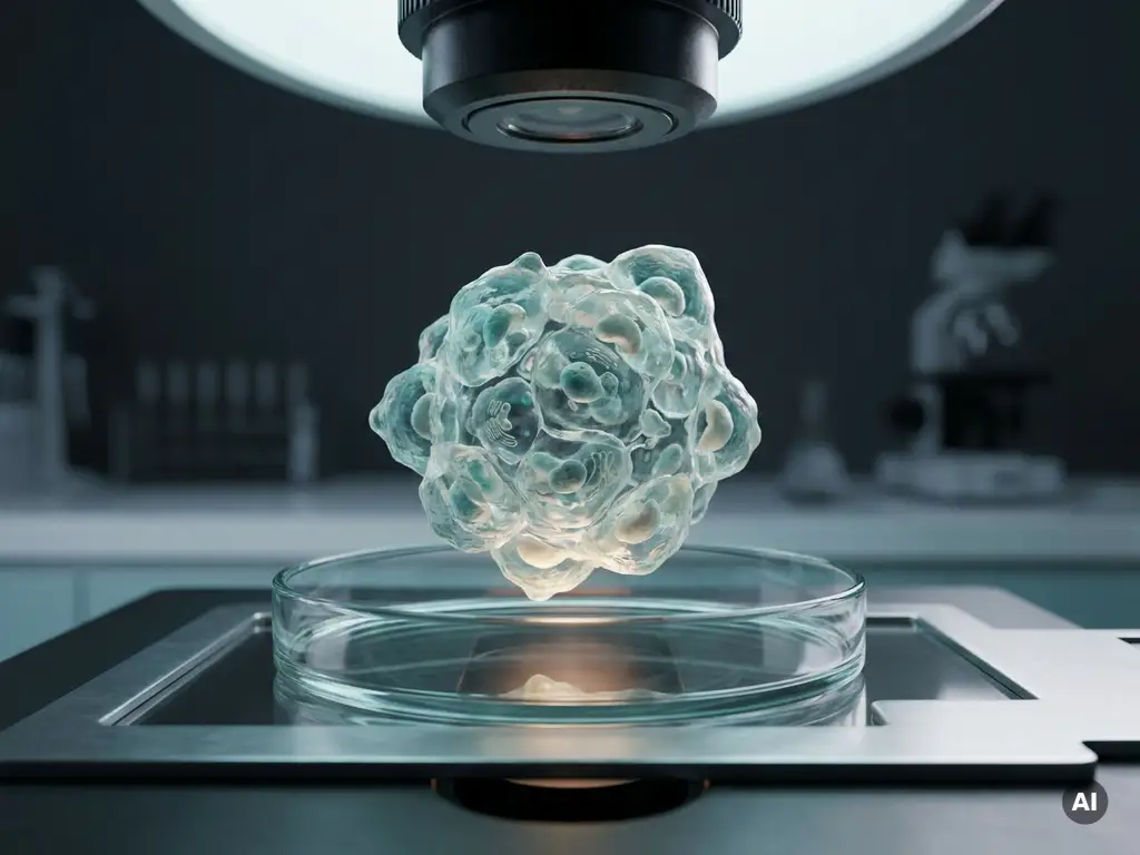
Label-free cell analysis is a collection of techniques that measure cell behavior, morphology, and physiology without the use of fluorescent dyes, radioactive tracers, or other exogenous labels. Instead of tagging cells with reporter molecules, these methods detect intrinsic cellular properties such as optical density, electrical impedance, refractive index, or mass. For oncology researchers, this approach enables long-term, minimally invasive observation of cancer cells under conditions that closely reflect their natural state. The sections below address the most common questions researchers in the Benelux region raise about this methodology.
How does label-free cell analysis actually work?
Label-free cell analysis works by detecting physical or biochemical properties inherent to cells rather than signals from added markers. Techniques such as impedance-based sensing, surface plasmon resonance, and quantitative phase imaging measure changes in electrical resistance, light refraction, or optical path length as cells grow, migrate, or respond to stimuli. Because no external label is introduced, the measurement reflects the cell’s own biology.
In impedance-based approaches, cells are seeded onto electrode arrays embedded in well plates. As cells attach, spread, and proliferate, they alter the flow of an alternating electrical current across the electrodes. The resulting impedance signal is recorded continuously and translated into a real-time readout of cell number, morphology, and adhesion strength.
Quantitative phase imaging and related label-free imaging techniques exploit the fact that cellular structures have a slightly different refractive index from the surrounding medium. By measuring how light waves are retarded as they pass through a cell, these systems can map dry mass distribution and structural organization at subcellular resolution without any staining step.
What types of cellular measurements can label-free methods capture?
Label-free methods can capture a broad range of cellular parameters, including proliferation, adhesion, migration, morphology, viability, and responses to chemical or mechanical stimuli. Because measurements are continuous rather than endpoint-based, researchers can observe dynamic processes such as cell cycle progression, cytoskeletal reorganization, and receptor-ligand interactions over hours or days within a single experiment.
Specific readouts available through different label-free platforms include:
- Cell index or normalized cell index from impedance sensors, reflecting overall cell health and attachment
- Optical path length and dry mass from quantitative phase imaging, indicating biomass accumulation
- Refractive index mapping for intracellular structural detail
- Binding kinetics from biosensor arrays, capturing receptor engagement and molecular interaction rates
- Migration velocity and directionality derived from time-lapse label-free imaging sequences
- Cytotoxicity profiles measured as impedance decline following compound addition
This breadth of readouts makes label-free cell analysis particularly suitable for multiparametric studies where a single assay needs to deliver mechanistic insight alongside quantitative data.
How does label-free analysis compare to fluorescence-based cell assays?
The key distinction is that fluorescence-based assays require the introduction of dyes, antibodies, or genetically encoded reporters, whereas label-free analysis relies entirely on intrinsic cellular signals. This difference has practical consequences for assay design, data quality, and the types of questions each approach can address.
Fluorescence-based methods offer high molecular specificity. By targeting a particular protein or organelle with a fluorescent probe, researchers can localize and quantify that specific molecule with precision. However, the introduction of labels carries risks: phototoxicity from repeated excitation, photobleaching over long time courses, and potential interference with the very biological processes under investigation.
Label-free analysis avoids these confounds entirely. Cells are not stressed by dye loading or light exposure at excitation wavelengths, making continuous live-cell imaging over extended periods far more practical. The trade-off is that label-free readouts are typically integrative rather than molecularly specific. An impedance signal, for example, reflects the combined contribution of cell number, shape, and adhesion rather than pinpointing a single molecular target.
In practice, many research groups in the Benelux combine both approaches within the same experimental workflow, using label-free monitoring to track overall cellular responses and then applying targeted fluorescence assays at selected time points to add molecular resolution where needed.
Why is label-free cell analysis particularly suited to oncology research?
Label-free cell analysis is particularly well suited to oncology research because cancer cell biology involves continuous, dynamic processes that endpoint assays capture poorly. Tumor cells proliferate, invade surrounding tissue, evade immune responses, and adapt to drug pressure over time. Label-free platforms can monitor all of these behaviors continuously without perturbing the cells, providing a more complete picture of how cancer cells behave under experimental conditions.
In the context of cancer cell analysis and drug screening, label-free biosensors allow researchers to observe cytotoxic responses in real time rather than at a fixed endpoint. This is important because some compounds may slow proliferation transiently before cells recover, a dynamic that a single-point viability assay would miss entirely.
Label-free analysis in oncology research also supports the study of tumor microenvironment interactions. Co-culture models, where cancer cells are grown alongside stromal or immune cells, can be monitored without the need to spectrally separate multiple fluorescent labels, simplifying experimental design and reducing the risk of signal crosstalk.
Additionally, because label-free methods are non-destructive, the same cell population can be analyzed repeatedly across a time course and then harvested for downstream molecular analysis. This is particularly valuable in studies using primary tumor material or patient-derived organoids, where sample quantities are limited.
What instruments and platforms are used for label-free cell analysis?
Several distinct instrument categories are used for label-free cell analysis, each based on a different physical detection principle and suited to different experimental questions in cancer cell biology and cell-based assays for oncology.
The main platform categories include:
- Impedance-based cell analyzers that use microelectrode well plates to continuously monitor cell attachment, proliferation, and cytotoxicity in real time
- Quantitative phase imaging systems that measure optical path length differences to map cell morphology and dry mass without staining
- Surface plasmon resonance instruments that detect molecular binding events at sensor chip surfaces, used for kinetic characterization of tumor-relevant receptor interactions
- Holographic and interferometric microscopes that reconstruct three-dimensional cell structure from phase information, enabling label-free live-cell imaging at high resolution
- Microfluidic biosensor platforms that integrate label-free detection with controlled flow conditions, supporting studies of cell migration and invasion under physiological shear stress
The choice of platform depends on the specific research question. Impedance systems are widely used in drug screening workflows because they integrate readily into standard multiwell plate formats. Quantitative phase and holographic imaging systems are preferred when spatial information about cell morphology or intracellular organization is required.
What are the limitations of label-free cell analysis in cancer studies?
The primary limitation of label-free cell analysis in cancer studies is reduced molecular specificity. Because these methods detect aggregate physical signals rather than labeled targets, it can be difficult to attribute a measured change to a specific molecular event. An impedance decrease, for instance, may reflect cell death, detachment, or morphological rounding, and distinguishing between these possibilities often requires additional assays.
Further limitations researchers should consider include:
- Signal complexity in co-culture models: When multiple cell types are present, the label-free signal reflects contributions from all cells simultaneously, making it challenging to isolate the behavior of a specific population
- Throughput constraints: Some label-free imaging platforms are optimized for depth of analysis rather than speed, which can limit their utility in high-throughput screening campaigns
- Sensitivity to non-biological variables: Temperature fluctuations, evaporation, and mechanical vibration can all influence impedance or optical measurements, requiring careful environmental control
- Limited compatibility with three-dimensional models: Many label-free biosensor formats were designed for adherent monolayer cultures and may not translate directly to spheroid or organoid models without platform-specific adaptations
Despite these constraints, label-free analysis remains a valuable component of the oncology research toolkit when used alongside complementary methods. Its strengths in continuous, non-invasive monitoring make it especially powerful for longitudinal studies and for experiments where preserving cell viability across the full time course is essential.
How BioSPX supports label-free cell analysis in the Benelux
BioSPX works with research laboratories, biotech companies, and pharmaceutical R&D teams across Belgium, the Netherlands, and Luxembourg to identify and implement label-free analysis solutions that fit their specific workflows. Whether a research group is establishing a new cell-based assay platform or expanding an existing oncology research program, BioSPX provides:
- Expert guidance on instrument selection based on experimental requirements and throughput needs
- Hands-on installation and configuration by a dedicated service team
- User training tailored to the research context, from assay development to data interpretation
- Ongoing maintenance support to ensure instrument performance and data reproducibility
- Specialist advice on integrating label-free platforms with broader laboratory workflows, including automation and liquid handling
If you are evaluating label-free cell analysis instruments for your oncology or life science research, contact the BioSPX team to discuss your requirements with a product specialist.
Frequently Asked Questions
How do I know which label-free platform is the right starting point for my oncology lab?
The best starting point depends on whether your primary need is functional monitoring or structural imaging. If you are running drug screening workflows with standard multiwell plates, an impedance-based system is typically the most practical first instrument because it integrates directly into existing plate-based formats and delivers continuous cytotoxicity and proliferation data with minimal protocol changes. If your work centers on morphological characterization or intracellular organization, a quantitative phase imaging system will deliver more spatial detail. Speaking with a specialist who understands both your research questions and your current infrastructure — such as the BioSPX team — can help you avoid investing in a platform that fits the technology but not the workflow.
Can label-free cell analysis be used with patient-derived tumor samples or organoids, or is it mainly suited to established cell lines?
Label-free analysis can be applied to patient-derived samples and organoids, but it requires careful consideration of platform compatibility. Many impedance-based systems are optimized for adherent monolayer cultures, so three-dimensional models such as tumor spheroids or organoids may need platform-specific adaptations, specialized plate formats, or complementary imaging approaches. The non-destructive nature of label-free monitoring is a genuine advantage here, since precious primary material can be observed continuously over the full experiment and then recovered for downstream molecular or genomic analysis. If you are working with organoid models, it is worth confirming with your instrument supplier whether validated protocols exist for your specific model system.
What are the most common mistakes researchers make when setting up a label-free impedance assay for the first time?
The most frequent errors relate to cell seeding density and environmental control. Seeding too many cells at the outset compresses the dynamic range of the assay, making it difficult to detect meaningful differences between treatment conditions; seeding too few cells delays attachment and increases well-to-well variability. Environmental factors such as temperature fluctuations, CO₂ instability, and evaporation from edge wells can all introduce noise into impedance signals that may be mistaken for a biological effect. Establishing a robust baseline period before adding compounds — typically 18 to 24 hours post-seeding — and running the instrument inside a standard cell culture incubator are two straightforward steps that significantly improve data quality from the outset.
How should I interpret a drop in impedance signal — does it always mean cells are dying?
A drop in impedance signal does not exclusively indicate cell death, which is one of the reasons complementary assays are valuable. Impedance decreases can reflect cell detachment, morphological rounding, cytoskeletal reorganization, or a reduction in cell-substrate contact area, all of which may occur independently of cytotoxicity. For example, a compound that disrupts actin dynamics may cause cells to round up and reduce their electrode coverage without immediately compromising viability. To distinguish between these possibilities, it is useful to correlate impedance data with orthogonal readouts such as a brightfield image at the time of the signal change, or a viability stain applied at a specific endpoint, to confirm the biological interpretation.
Is it feasible to run label-free analysis and fluorescence-based assays simultaneously on the same cell population?
In many cases, yes — and this combination is increasingly common in oncology research groups. Some impedance platforms are designed to be compatible with fluorescence microscopy, allowing researchers to overlay impedance kinetics with fluorescence images captured at selected time points without transferring cells to a different vessel. Quantitative phase imaging systems can also be paired with low-intensity fluorescence channels on compatible microscope platforms, provided the excitation light dose is kept low enough to avoid phototoxicity. The practical benefit is that you retain the continuous, non-invasive monitoring of label-free analysis while gaining the molecular specificity of a targeted fluorescence assay at the moments in the time course where mechanistic detail matters most.
How much technical expertise is required to get meaningful data from label-free platforms, and is there a steep learning curve?
The core operation of impedance-based systems is relatively accessible — seeding cells into electrode plates and initiating a run requires skills comparable to a standard cell viability assay, and most platforms provide software that automates data acquisition and generates real-time curves without manual intervention. The steeper learning curve lies in data interpretation: understanding how to design the right controls, recognize artefactual signal changes, and extract biologically meaningful conclusions from multiparametric readouts takes time and ideally some guided experience. Quantitative phase imaging systems have a higher initial setup complexity, particularly around optical alignment and data analysis pipelines. Structured user training at the point of instrument installation — covering both assay development and data interpretation — substantially shortens the time to reliable, publication-quality results.
Can label-free analysis support regulatory or preclinical drug development workflows, or is it primarily a research discovery tool?
Label-free analysis is increasingly used in preclinical drug development contexts, particularly for early-stage compound profiling, mechanism-of-action studies, and cytotoxicity screening, where its continuous real-time readout provides richer pharmacodynamic information than endpoint assays alone. Impedance-based cytotoxicity data, for example, can reveal whether a compound’s effect is cytostatic or cytotoxic, and at what concentration and time point the response becomes irreversible — all of which are relevant to preclinical candidate selection. Regulatory acceptance of label-free data as a standalone submission-ready readout is still evolving, so in validated GxP workflows these methods are typically used alongside established endpoint assays rather than as a direct replacement. Discussing your specific regulatory context with your instrument provider early in assay development will help ensure the data you generate meets the documentation and reproducibility standards required downstream.
Related Articles
- Why is spatial context important for understanding molecular mechanisms?
- How do researchers distinguish cytostatic from cytotoxic drug effects in vitro?
- What causes genomic instability in cancer cells?
- What are the most significant advances in oncology in 2026?
- How do genetic mutations contribute to cancer development?
This content was generated with the help of AI and it may contain mistakes