
 EN
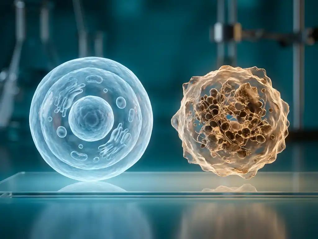
Cancer cells differ from normal healthy cells in several fundamental ways: they grow and divide without control, ignore signals to stop, invade surrounding tissues, and can spread to distant organs. These differences arise from accumulated genetic mutations that disrupt the normal regulatory mechanisms governing cell behavior. Understanding these distinctions is central to cancer biology research and to developing better tools for studying tumor cells in the laboratory.
The sections below unpack the most commonly asked questions about how cancer cells behave differently from normal cells, from their metabolic strategies to their microscopic appearance.
Why do cancer cells grow and divide uncontrollably?
Cancer cells grow and divide uncontrollably because mutations in key regulatory genes disrupt the normal checkpoints that govern the cell cycle. In healthy cells, a tightly regulated system of signals determines when a cell should divide, pause, or stop. Cancer cells bypass these controls, enabling continuous proliferation even in the absence of the growth signals that normal cells require.
Two categories of genes are most frequently involved. Proto-oncogenes, when mutated into oncogenes, act like a stuck accelerator, driving cell division regardless of external signals. Tumor suppressor genes, such as TP53 and RB1, normally function as brakes on cell division. When these are inactivated by mutation, the cell loses its ability to pause or self-correct.
Cancer cells also acquire the ability to sustain their own growth signals, a property researchers describe as self-sufficiency in growth signaling. Rather than waiting for external cues, they produce their own stimulatory molecules, creating a self-reinforcing loop of uncontrolled division.
What makes cancer cells able to ignore signals to stop growing?
Cancer cells are able to ignore stop signals because they develop resistance to growth-inhibitory cues, a hallmark of malignant transformation. In normal tissue, neighboring cells and the surrounding extracellular matrix send inhibitory signals that prevent overcrowding and maintain tissue architecture. Tumor cells acquire mutations that make them unresponsive to these signals.
One well-studied mechanism involves the loss of contact inhibition. Healthy cells stop dividing when they come into physical contact with surrounding cells. Cancer cells lose this sensitivity, allowing them to pile up and form tumors.
Additionally, cancer cells frequently inactivate apoptosis, the programmed cell death pathway that eliminates damaged or abnormal cells. By overexpressing anti-apoptotic proteins or disabling pro-apoptotic ones, tumor cells effectively become resistant to the molecular instructions that would normally trigger their elimination. This evasion of cell death is as important to tumor growth as the acceleration of cell division itself.
How do cancer cells look different from normal cells under a microscope?
Under a microscope, cancer cells display a range of structural abnormalities that distinguish them clearly from normal healthy cells. These morphological differences are the basis of histopathological diagnosis and remain one of the most reliable indicators of malignancy in research and clinical settings.
Key observable differences include:
- Enlarged and irregular nuclei: Cancer cell nuclei are often disproportionately large relative to the cytoplasm, with an abnormal nuclear-to-cytoplasmic ratio.
- Prominent nucleoli: The nucleoli, structures within the nucleus involved in ribosome production, are frequently enlarged and more visible in tumor cells.
- Pleomorphism: Cancer cells vary significantly in size and shape, unlike the uniform appearance of normal cells in a tissue.
- Abnormal mitotic figures: Dividing cancer cells often show irregular or multipolar mitotic spindles, reflecting chromosomal instability.
- Loss of tissue organization: Normal cells arrange themselves in organized layers or structures. Cancer cells lose this ordered architecture, appearing disorganized under the microscope.
These visual characteristics are studied extensively in laboratory research using imaging systems and microscopy platforms, which allow researchers to analyze cell morphology at high resolution and track changes over time.
Automated imaging platforms such as the BioTek Lionheart FX capture these morphological features across brightfield, phase contrast, and multi-channel fluorescence, allowing researchers to quantify nuclear-to-cytoplasmic ratio, mitotic abnormalities, and pleomorphism consistently across large cell populations rather than relying on manual scoring.
What is the Warburg effect and why do cancer cells use it?
The Warburg effect is the observation that cancer cells preferentially use glycolysis to produce energy, even in the presence of sufficient oxygen, rather than the more efficient oxidative phosphorylation pathway used by normal cells. This phenomenon, first described by Otto Warburg in the early twentieth century, remains one of the most studied metabolic hallmarks of cancer.
Normal cells rely on oxidative phosphorylation in the mitochondria when oxygen is available, generating far more ATP per glucose molecule than glycolysis. Cancer cells, however, favor aerobic glycolysis despite its lower energy yield. This appears counterintuitive, but research suggests the strategy offers several advantages for rapidly dividing cells.
Glycolysis produces metabolic intermediates that feed into biosynthetic pathways, supplying the building blocks needed for rapid cell proliferation, including nucleotides, amino acids, and lipids. The high rate of glucose consumption also acidifies the tumor microenvironment, which can suppress immune responses and facilitate invasion of surrounding tissue. The Warburg effect is actively investigated as a research target because it represents a metabolic vulnerability that distinguishes tumor cells from most normal tissues.
How do cancer cells spread to other parts of the body?
Cancer cells spread to other parts of the body through a process called metastasis, which involves a sequence of steps that allow tumor cells to detach from the primary site, enter the bloodstream or lymphatic system, survive in circulation, and establish new tumors at distant locations.
This process unfolds in several stages:
- Local invasion: Cancer cells degrade the extracellular matrix surrounding the tumor using enzymes such as matrix metalloproteinases, allowing them to invade adjacent tissue.
- Intravasation: Tumor cells penetrate blood vessel or lymphatic walls and enter circulation as circulating tumor cells.
- Survival in circulation: Most circulating tumor cells are destroyed by physical forces or immune cells, but some survive by aggregating with platelets or evading immune detection.
- Extravasation: Surviving cells exit the bloodstream at a distant site by migrating through vessel walls.
- Colonization: Cells establish themselves in a new tissue environment, a step that requires adaptation to a foreign microenvironment and is considered a major bottleneck in metastasis.
A key molecular driver of metastasis is the epithelial-to-mesenchymal transition (EMT), a process in which cancer cells lose their adhesive properties and gain migratory characteristics. This transition is an active area of cancer biology research, as it also contributes to resistance to certain research interventions. Instruments such as the Agilent xCELLigence RTCA DP extend this further by continuously tracking invasion and migration in real time through an integrated Boyden chamber, capturing the full kinetics of EMT-driven behavior rather than relying on scratch-assay endpoints alone.
Can cancer cells ever revert to behaving like normal cells?
Under certain experimental conditions, cancer cells can be induced to revert toward more normal behavior, a phenomenon known as cancer cell reprogramming or differentiation. However, this is not a spontaneous or common occurrence, and the extent to which it is possible depends on the type of cancer and the specific mutations involved.
Research in cellular reprogramming has demonstrated that some tumor cells retain a degree of plasticity. Exposure to specific molecular signals or changes in the microenvironment can push cancer cells toward a more differentiated, less proliferative state. In some experimental models, highly malignant cells have been reprogrammed into cells that more closely resemble their tissue of origin.
This area of investigation is considered promising in cancer biology research because it raises the possibility of targeting the mechanisms that maintain the undifferentiated, aggressive state of tumor cells. The concept connects closely to research on cancer stem cells, which are thought to drive tumor maintenance and recurrence and are currently being studied as a focus of laboratory research in the Benelux region and beyond.
How BioSPX supports cancer biology research
Studying the differences between cancer cells and normal healthy cells requires precise, reliable laboratory instrumentation. Researchers investigating tumor cell behavior, metabolic changes, metastatic mechanisms, and cellular morphology depend on high-performance tools that deliver reproducible results.
BioSPX supports life science researchers across the Benelux region with an extensive portfolio of instruments suited to cancer biology workflows, including:
- Imaging and microscopy systems for analyzing cancer cell morphology, migration, and tissue architecture
- Multimode readers for assessing cell viability, metabolic activity, and proliferation assays
- Liquid handling solutions for high-throughput and reproducible experimental workflows
- Cell culture automation systems such as the Nestor, which automates routine tasks for up to 10 flasks or multiwell plates, reducing variability and freeing researchers to focus on analysis
- Organoid research solutions including the Orgonex RPMotion bioreactor, supporting three-dimensional tumor model research
BioSPX’s product specialists provide expert guidance on instrument selection and experimental setup, while the service department ensures correct installation and rapid maintenance response. If you are setting up or scaling a cancer biology research workflow in Belgium, the Netherlands, or Luxembourg, contact BioSPX to discuss which solutions best fit your research needs.
Frequently Asked Questions
What is the best starting point for setting up a cancer cell biology lab workflow?
The most practical starting point is to define the specific biological questions you are investigating — whether that is cell proliferation, morphology, metabolism, or metastatic behavior — and then select instruments that address those assays reproducibly. For most cancer biology workflows, a combination of a multimode plate reader for viability and metabolic assays, an imaging system for morphological analysis, and a cell culture automation solution to minimize manual variability provides a strong foundation. Consulting with a product specialist familiar with cancer research workflows, such as those at BioSPX, can help you avoid costly mismatches between your experimental needs and instrument capabilities.
How do researchers distinguish between cancer cells and normal cells in a mixed culture?
Researchers typically use a combination of morphological analysis, molecular markers, and functional assays to distinguish cancer cells from normal cells in co-culture systems. Fluorescent labeling with cell-type-specific antibodies or reporter gene constructs allows live-cell imaging systems to track each population separately. Flow cytometry is another widely used approach, enabling quantitative separation of cell populations based on surface marker expression. For studies where spatial context matters — such as invasion assays or organoid models — high-resolution imaging platforms that capture both morphology and fluorescence simultaneously are particularly valuable.
Are 2D cell culture models sufficient for studying cancer cell behavior, or are 3D models necessary?
Traditional 2D monolayer cultures remain useful for many standardized assays, but they fail to replicate the architecture, oxygen gradients, nutrient distribution, and cell-cell interactions found in actual tumors. Three-dimensional models such as spheroids and organoids more accurately recapitulate tumor microenvironment conditions, making them increasingly preferred for studies of invasion, drug response, and metastatic behavior. For researchers transitioning to 3D workflows, dedicated bioreactor systems — such as those designed for organoid culture — provide the controlled dynamic environment these models require to develop reproducibly and at scale.
What are the most common mistakes researchers make when working with cancer cell lines in the lab?
One of the most frequent and consequential mistakes is failing to authenticate cell lines regularly, which can lead to years of research built on misidentified or cross-contaminated cultures. Short tandem repeat (STR) profiling is the standard method for confirming cell line identity and should be performed at regular intervals. Other common pitfalls include allowing cells to drift in phenotype through excessive passaging, inconsistent culture conditions that introduce variability between experiments, and neglecting mycoplasma testing. Automating routine culture tasks with cell culture automation systems helps reduce human-introduced variability and improves reproducibility across experiments.
How is the Warburg effect exploited in cancer research workflows, and what assays are used to measure it?
Researchers exploit the Warburg effect by using metabolic assays that measure glucose consumption, lactate production, and oxygen consumption rates — all of which reflect the balance between glycolysis and oxidative phosphorylation in a cell population. Multimode plate readers equipped with fluorescence and luminescence detection capabilities are commonly used to run these metabolic activity assays in a high-throughput format. Comparing the metabolic profiles of cancer cells versus normal counterparts under the same conditions can reveal metabolic vulnerabilities that may be relevant to experimental intervention strategies.
Can the epithelial-to-mesenchymal transition (EMT) be observed and quantified in the laboratory?
Yes, EMT can be tracked using a combination of molecular and imaging-based approaches. At the molecular level, researchers monitor the loss of epithelial markers such as E-cadherin and the gain of mesenchymal markers such as vimentin and N-cadherin using immunofluorescence, Western blot, or gene expression assays. Functionally, EMT is often assessed through scratch assays, transwell migration assays, or invasion assays, all of which can be imaged and quantified using automated imaging systems. Longitudinal live-cell imaging is particularly informative, as it captures the dynamic nature of the transition rather than providing only a single time-point snapshot.
How do cancer stem cells differ from the bulk tumor cell population, and why does this matter for research?
Cancer stem cells (CSCs) represent a subpopulation within a tumor that exhibits enhanced self-renewal capacity, resistance to many experimental perturbations, and the ability to regenerate the heterogeneous cell population of the original tumor. Unlike the bulk of tumor cells, CSCs are thought to drive tumor recurrence and long-term maintenance, which is why they are an increasingly important focus of cancer biology research. Identifying and isolating CSCs typically relies on specific surface markers and functional assays such as sphere-forming efficiency in non-adherent culture conditions. Three-dimensional organoid models are particularly well suited for studying CSC behavior, as they preserve the hierarchical organization and microenvironmental cues that influence stem cell activity within tumors.
Related Articles
- How does FRET measure molecular proximity below the resolution limit of conventional microscopy?
- How can image segmentation improve quantitative spatial biology analysis?
- How does tumor spheroid formation differ from monolayer cell culture?
- What can continuous cell monitoring reveal that endpoint assays cannot?
- What is oncology and what does it involve?
This content was generated with the help of AI and it may contain mistakes