BioTek Lionheart FX
Information
Lionheart™ FX Automated Microscope is a compact, inclusive microscopy system for a broad range of imaging workflows.
It offers up to 100x air and oil immersion magnification, with:
- Fluorescence,
- Brightfield,
- Color brightfield,
- and phase contrast channels for maximum application reach.
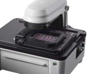 An optional environment control cover provides incubation to 40°C and effective containment for CO2/O2 control, a humidity chamber optimizes conditions for long-term live cell imaging applications, and an available dual reagent injector facilitates rapid kinetic assays. Lionheart FX and Gen5 together comprise Augmented Microscopy™ – the automation of image capture, processing, analysis and development of publication-ready images and data.
-
Lionheart FX vs Lionheart LX
Lionheart FX Lionheart LX General Microplate types 6- to 1536-well plates 6- to 1536-well plates Other labware supported Microscope slides, Petri and cell culture dishes, cell culture flasks (T25, T75), counting chambers (hemocytometers), chamber slides Microscope slides, Petri and cell culture dishes, cell culture flasks (T25, T75), counting chambers (hemocytometers), chamber slides Support for labware up to 1.5″ tall Support for labware up to 1.5″ tall Temperature control Incubation to 40 °C with optional environmental control cover Software Gen5 Microplate Reader and Imager Software included Gen5 Microplate Reader and Imager Software included Gen5 Image and Image+ Software available for advanced image analysis (option) Gen5 Image and Image+ Software available for advanced image analysis (option) CO2 and O2 control (option) 0 – 20% CO2 control and 1 – 19% O2 control, with optional Gas Controller Environmental control cover Top cover for light tight imaging and incubation control (option) X/Y Stage resolution Lead screw driven stage with 0.1 micron resolution Lead screw driven stage with 0.1 micron resolution Humidity control Humidity chamber with rapid gas recharge (option) Physical Characteristics Power 250 Watts max. 250 Watts max. Dimensions With cover closed or without cover: 18.3″ D, 17.9″ W, 14.1″ H (46.5 cm x 45.5 cm x 35.8 cm) 18.3″ D, 17.9″ W, 14.1″ H (46.5 cm x 45.5 cm x 35.8 cm) With cover fully open: 18.3″ D, 17.9″ W, 27.5″ H (46.5 cm x 45.5 cm x 69.8 cm) Weight Without environmental control cover: 51 lbs (23.1 kg) 51 lbs (23.1 kg) With environmental control cover: 58 lbs (26.3 kg) Regulatory Regulatory CE and TUV marked. RoHS compliant. Models for In Vitro Diagnostic use are available. CE and TUV marked. RoHS compliant. Models for In Vitro Diagnostic use are available. Imaging System Imaging mode Fluorescence, brightfield, color brightfield, and phase contrast Fluorescence, high contrast brightfield and color brightfield Imaging method Single color, multi-color, montage, time lapse, Z-stacking, burst mode Single color, multi-color, montage, time lapse, Z-stack, z-stack montage, burst mode Light source High power LEDs (available wavelengths: 365 nm, 390 nm, 465 nm, 505 nm, 523 nm, 590 nm, 623 nm, 655 nm, 740 nm) High power LEDs (available wavelengths: 365 nm, 390 nm, 465 nm, 505 nm, 523 nm, 590 nm, 623 nm, 655 nm, 740 nm) Camera 16-bit gray scale, Sony CCD, 1.25 megapixel 16-bit CMOS camera. 2.3 MP Sony IMX249 1/1.2″ sensor with 5.86 micron pixel size Camera binning Optional 2×2 binning for focus and/or image capture Optional 2×2 binning for focus and/or image capture Camera exposure range 5 milliseconds to 4 seconds 5 milliseconds to 4 seconds Image outputs available Raw Images: 16-bit TIFF Raw Images: 16-bit TIFF Saved Images: TIF, JPG, BMP, PNG, EMF, GIF Saved Images: TIFF, JPG, BMP, PNG, EMF, GIF, color TIFF Movies: MP4, WMV Movies: MP4, WMV Objective capacity 6 onboard, user-replaceable objectives 6 onboard, user-replaceable objectives Objectives available Fluorescence: Air: 1.25x, NA .04; 2.5x (2.25x eff), NA: .07; 2.5x (2.75x eff), NA: .12; 4x, NA: .13; 10x, NA: .30; 20x, NA: .45; 40x, NA.60; 60x, NA .70 Air: 1.25x, NA .04; 2.5x (2.25x eff), NA: .07; 2.5x (2.75x eff), NA: .12; 4x, NA: .13; 10x, NA: .30; Oil: 60x, NA:1.42; 60x, NA 1.25; 100x, NA 1.4; 100x, NA 1.3 20x, NA: .45; 40x, NA.60; 60x, NA .70 High NA: 20x, NA 0.75; 40x, NA 0.95 Oil: 60x, NA:1.42; 100x, NA: 1.40 Phase objectives available: 4x, NA: .13; 10x, NA: .30; 20x, NA: .45; 40x, NA .60 Image filter cube capacity 4 user-replaceable fluorescence cubes plus brightfield channel 4 user-replaceable fluorescence cubes plus brightfield channel Imaging filter cubes available DAPI, CFP, GFP, YFP, RFP, Texas Red, CY5, CY7, Acridine Orange, CFP-YFP FRET, Chlorophyll, Phycoerythrin (PE), Propidium Iodide, CY5.5, TagBFP, GFP (Ex)-CY6 (Em), RFP (Ex)-CY5 (Em), Alexa 568, Ex377 / Em647 DAPI, CFP, GFP, YFP, RFP, Texas Red, CY5, CY7, Acridine Orange, CFP-YFP FRET, Chlorophyll, Phycoerythrin (PE), Propidium Iodide, CY5.5, TagBFP, GFP (Ex)-CY6 (Em), RFP (Ex)-CY5 (Em), Alexa 568, Ex377 / Em647 Automated functions Autofocus, user-trained autofocus, auto exposure, auto-LED intensity Autofocus, user-trained autofocus, auto exposure, auto-LED intensity Autofocus method Image-based autofocus Image-based autofocus Laser autofocus option Laser autofocus option Image collection rate Single well fastest frame rate capture: Single well fastest frame rate capture: *Full Resolution: up to 10 frames per second for single color images *Full Resolution: up to 10 frames per second for single color images *2×2 Binning: up to 20 frames per second for single color images *2×2 Binning: up to 20 frames per second for single color images Microscope stage control Gen5 Software Control Gen5 Software Control Optional joystick controller Optional joystick controller Reagent Injectors (option) Number 2 syringe pumps Supported labware 6- to 384-well microplates, Petri and cell culture dishes, chamber slides Dead volume <1.65 mL with back flush Dispense tip options Aligned tip – aligned with optical path for dispensing for fast kinetic assays Offset tip – dispensing is offset from the optical path Dispense volume 5 – 1000 µL in 1 µL increments Dispense accuracy +1 µL or 2% Dispense precision <2% at 50 – 200 µL Specifications are subject to change. -
Specifications
GENERAL Microplate types 6- to 1536-well plates Other labware supported Microscope slides, Petri and cell culture dishes, cell culture flasks (T25, T75), counting chambers (hemocytometers), chamber slides Support for labware up to 1.5″ tall Temperature control Incubation to 40 °C with optional environmental control cover Software Gen5 Microplate Reader and Imager Software included Gen5 Image and Image+ Software available for advanced image analysis (option) CO2 and O2 control (option) 0 – 20% CO2 control and 1 – 19% O2 control, with optional Gas Controller Environmental control cover Top cover for light tight imaging and incubation control (option) X/Y Stage resolution Lead screw driven stage with 0.1 micron resolution Humidity control Humidity chamber with rapid gas recharge (option) IMAGING SYSTEM Imaging mode Fluorescence, brightfield, color brightfield, and phase contrast Imaging method Single color, multi-color, montage, time lapse, Z-stacking, burst mode Light source High power LEDs (available wavelengths: 365 nm, 390 nm, 465 nm, 505 nm, 523 nm, 590 nm, 623 nm, 655 nm, 740 nm) Camera 16-bit gray scale, Sony CCD, 1.25 megapixel Camera binning Optional 2×2 binning for focus and/or image capture Camera exposure range 5 milliseconds to 4 seconds Image outputs available Raw Images: 16-bit TIFF Saved Images: TIF, JPG, BMP, PNG, EMF, GIF Movies: MP4, WMV Objective capacity 6 onboard, user-replaceable objectives Objectives available Fluorescence: Air: 1.25x, NA .04; 2.5x (2.25x eff), NA: .07; 2.5x (2.75x eff), NA: .12; 4x, NA: .13; 10x, NA: .30; 20x, NA: .45; 40x, NA.60; 60x, NA .70 Oil: 60x, NA:1.42; 100x, NA: 1.40 Phase objectives available: 4x, NA: .13; 10x, NA: .30; 20x, NA: .45; 40x, NA .60 Image filter cube capacity 4 user-replaceable fluorescence cubes plus brightfield channel Imaging filter cubes available DAPI, CFP, GFP, YFP, RFP, Texas Red, CY5, CY7, Acridine Orange, CFP-YFP FRET, Chlorophyll, Phycoerythrin (PE), Propidium Iodide, CY5.5, TagBFP, GFP (Ex)-CY6 (Em), RFP (Ex)-CY5 (Em), Alexa 568, Ex377 / Em647 Automated functions Autofocus, user-trained autofocus, auto exposure, auto-LED intensity Autofocus method Image-based autofocus Laser autofocus option Image collection rate Single well fastest frame rate capture: *Full Resolution: up to 10 frames per second for single color images *2×2 Binning: up to 20 frames per second for single color images Microscope stage control Gen5 Software Control Optional joystick controller REAGENT INJECTORS (OPTION) Number 2 syringe pumps Supported labware 6- to 384-well microplates, Petri and cell culture dishes, chamber slides Dead volume <1.65 mL with back flush Dispense tip options Aligned tip – aligned with optical path for dispensing for fast kinetic assays Offset tip – dispensing is offset from the optical path Dispense volume 5 – 1000 µL in 1 µL increments Dispense accuracy +1 µL or 2% Dispense precision <2% at 50 – 200 µL PHYSICAL CHARACTERISTICS Power 250 Watts max. Dimensions With cover closed or without cover: 18.3″ D, 17.9″ W, 14.1″ H (46.5 cm x 45.5 cm x 35.8 cm) With cover fully open: 18.3″ D, 17.9″ W, 27.5″ H (46.5 cm x 45.5 cm x 69.8 cm) Weight Without environmental control cover: 51 lbs (23.1 kg) With environmental control cover: 58 lbs (26.3 kg) REGULATORY Regulatory CE and TUV marked. RoHS compliant. Models for In Vitro Diagnostic use are available. -
Features
Automated digital microscopy to 100x.
Lionheart FX with Augmented Microscopy automates make your imaging workflow to yield amazing images and powerful data. Automated image capture starts with image-based and laser autofocus, plus auto LED and exposure. Automated image pre-processing optimizes images for downstream analysis, from cell counting to characterization of subcellular details.Four imaging modes, multiple imaging processes
With brightfield, color brightfield, phase contrast and fluorescence imaging in four channels – and nearly twenty available filter/LED cubes, Lionheart FX offers fantastic versatility that isn’t limited to image capture. Follow your imaging processes through z-stacking to z-projection, montage collection to stitching and digital phase contrast to analysis, all one hardware and software platform.Label-free cell counting
It’s fast and simple to count live cells using the available high contrast brightfield option. Higher contrast allows a direct and label-free method for object identification using Gen5 software to count cells and characterize cell proliferation in kinetic studies.Kinetic live cell assay support
Several options are available to support live cell assays with Lionheart FX, including an environmental cover to maintain temperature and gas levels within the system. Long-term live cell kinetics are supported, using the unique humidity chamber that fits on the stage itself, keeping the live cell in a completely nurturing environment. A dual reagent injector provides quick inject/image capability for observing fast reactions.Fully integrated and compact
Fifteen minutes is all it takes to install and set up Lionheart FX for image capture and analysis. The filter/LED cubes and objectives are accessed from the front panel, and tool-free connections for injectors, gas control and the environmental cover add to the easy installation. Its small size takes up minimal benchtop space. -
Configurations
LFX
Lionheart FX Automated Live Cell Imager. Fluorescence, brightfield, color brightfield and phase contrast imaging capabililty. Includes base Gen5 software. Objectives, filter/LED cubes, accessories and software upgrades sold separately.
Brochures
Application notes
Please login to be able to see the application notes.