
 EN
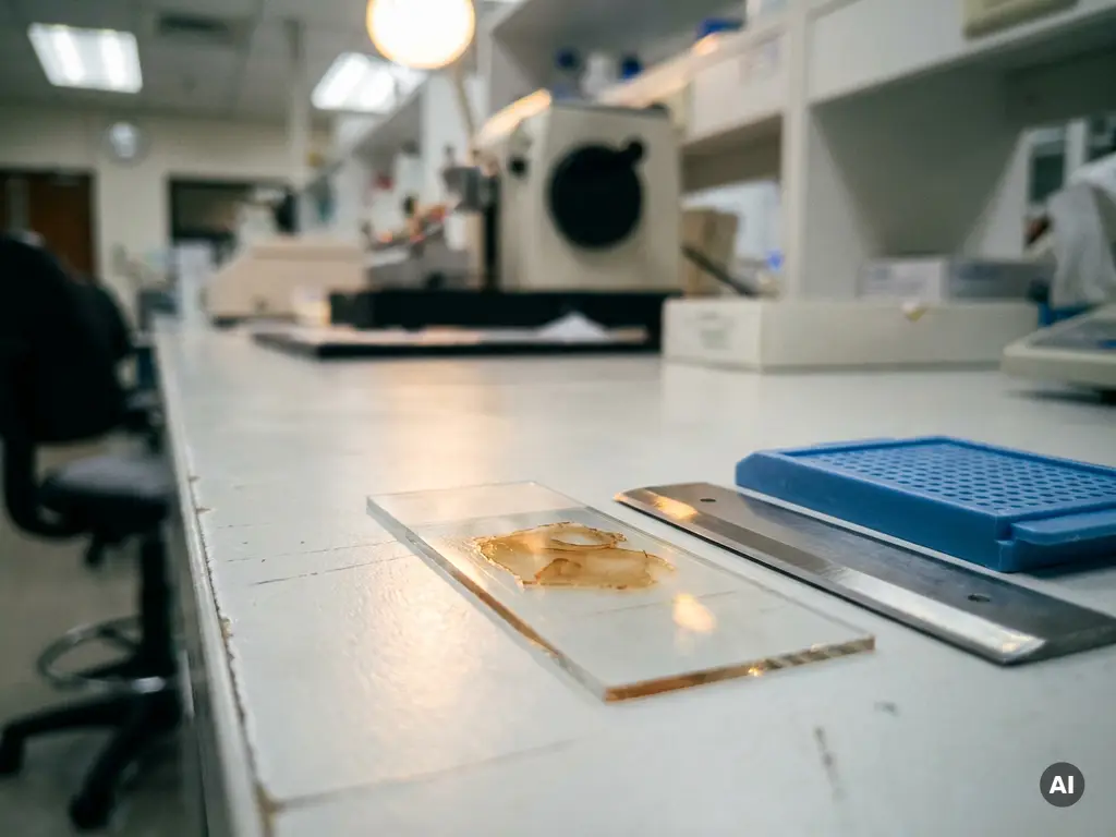
Researchers analyzing formalin-fixed paraffin-embedded tissue samples must account for chemical modifications introduced during fixation, variable nucleic acid and protein integrity, and the need for rigorous quality controls before downstream assays. FFPE tissue analysis presents unique challenges that differ substantially from fresh or frozen tissue, making careful sample preparation and method selection essential for reproducible results. The questions below address the most critical considerations at each stage of the workflow.
Why is FFPE tissue chemically challenging to work with?
FFPE tissue is chemically challenging because formalin fixation introduces extensive cross-links between proteins and nucleic acids, altering the molecular architecture of the sample. These cross-links preserve tissue morphology effectively, which is why formalin-fixed paraffin-embedded tissue has been the standard in pathology for decades, but they simultaneously fragment DNA and RNA and make proteins harder to extract in their native conformation.
Formaldehyde reacts with free amino groups in proteins and nucleic acids, forming methylene bridges that lock molecules together. Over time, these adducts can become increasingly stable, reducing the efficiency of downstream extraction and analysis. RNA is particularly susceptible, as it degrades more readily than DNA under fixation conditions and during subsequent storage. The result is a sample type that is molecularly compromised even when histologically well preserved.
For researchers in Benelux laboratories working with archival biobanks or retrospective cohort materials, this chemical complexity is a routine reality. Understanding the underlying chemistry is the first step toward selecting appropriate extraction protocols and analytical methods that can accommodate the limitations of the material.
How does deparaffinization affect downstream assay quality?
Deparaffinization, the removal of paraffin wax before molecular analysis, directly affects downstream assay quality by determining how completely and consistently the tissue is accessible for extraction. Incomplete deparaffinization leaves residual wax that can inhibit enzymatic reactions, reduce nucleic acid yields, and introduce variability between samples.
The standard approach involves xylene or xylene-substitute washes followed by a graded ethanol series to rehydrate the tissue. The efficiency of this process depends on section thickness, tissue type, and the age of the paraffin block. Thicker sections and older blocks tend to retain more wax, increasing the risk of inhibition in downstream steps such as PCR amplification or sequencing library preparation.
Residual paraffin can also interfere with protein-based assays, including immunohistochemistry and mass spectrometry-based proteomics, by blocking antibody access or co-eluting with target proteins. Researchers should validate their deparaffinization protocol on representative material before committing to a full analytical run, particularly when working with samples from different institutions or storage conditions.
What are the key considerations for extracting nucleic acids from FFPE samples?
Extracting nucleic acids from FFPE tissue samples requires balancing efficient cross-link reversal with minimizing additional fragmentation. The three most important considerations are cross-link reversal conditions, proteinase K digestion efficiency, and the selection of an extraction chemistry suited to the expected degree of molecular degradation.
- Cross-link reversal: Heat-induced reversal at elevated temperatures (typically 56 to 90 degrees Celsius) is the most widely used approach, but incubation time and temperature must be optimized. Excessive heat can cause further DNA fragmentation, while insufficient heat leaves cross-links intact and reduces yield.
- Proteinase K digestion: Thorough protein digestion is essential for releasing nucleic acids trapped within the cross-linked matrix. Digestion conditions should be titrated to the tissue type and fixation duration.
- Fragment size awareness: FFPE-derived DNA and RNA are typically fragmented to shorter lengths than fresh material. Extraction protocols and downstream assays should be selected with this in mind, favoring short amplicon designs in PCR and fragment-aware library preparation for sequencing.
- RNA integrity: FFPE RNA is often severely degraded. Assays relying on intact transcripts, such as standard RNA-seq, may require specialized low-input or degraded-RNA-compatible protocols.
- Inhibitor removal: Formalin-derived contaminants and other inhibitors co-purify with nucleic acids. Washing steps and column-based cleanup are important for achieving amplifiable extracts.
Researchers working in pharmaceutical R&D or academic settings across Belgium and the Netherlands frequently rely on FFPE-derived nucleic acids for biomarker discovery and retrospective studies. Validating extraction efficiency with quantitative and qualitative metrics before analysis is strongly recommended.
Which analytical techniques are best suited for FFPE-derived material?
The analytical techniques best suited for FFPE tissue samples are those designed to work with short, chemically modified, or low-abundance molecular targets. Technique selection should be guided by the degree of degradation in the sample and the biological question being asked.
For nucleic acid analysis, short-amplicon quantitative PCR is one of the most robust approaches, as it accommodates the fragmented nature of FFPE DNA and RNA. Next-generation sequencing panels optimized for FFPE material, including targeted gene panels and hybrid capture-based methods, can deliver reliable variant detection even from degraded input. Whole-genome or whole-transcriptome approaches are more challenging and typically require higher-quality input than most FFPE samples provide.
For protein and tissue biomarker analysis, immunohistochemistry remains a cornerstone technique because it preserves spatial context and works directly on fixed sections. Multiplex immunofluorescence extends this capability by enabling simultaneous detection of multiple markers within the same tissue section, which is valuable for understanding co-expression patterns in complex tissue environments. Mass spectrometry-based proteomics has also advanced significantly for FFPE applications, enabling quantitative protein profiling from archived material.
Researchers interested in cellular analysis workflows should consider how imaging-based techniques can complement molecular readouts, particularly when tissue architecture and cellular context are important to the research question.
How does tissue storage duration impact FFPE sample integrity?
Storage duration is one of the most significant variables affecting FFPE sample integrity. As archived blocks age, the chemical cross-links introduced during formalin fixation continue to evolve, nucleic acids undergo progressive oxidative damage, and protein epitopes may become increasingly masked or degraded.
DNA from blocks stored for fewer than five years generally yields more amplifiable material than DNA from blocks stored for a decade or longer, though this relationship is not strictly linear and varies with storage conditions. Temperature fluctuations, humidity, and light exposure all accelerate degradation. Sections cut from old blocks and left unmounted on slides deteriorate faster than intact paraffin blocks, making it preferable to cut sections as close to the time of analysis as possible.
RNA degradation is even more pronounced over time. Transcriptomic studies using archival FFPE material from biobanks with samples spanning many years should include storage duration as a covariate in experimental design and statistical analysis. Researchers should also be aware that fixation quality at the time of processing, including fixation time and formalin concentration, interacts with storage duration to determine the overall molecular quality of the sample.
What quality controls should be in place before analyzing FFPE samples?
Quality controls for FFPE tissue samples should be established before any downstream analysis begins, covering both the integrity of the extracted material and the performance of the analytical assay. Skipping or minimizing quality assessment at this stage is a leading cause of failed experiments and irreproducible results.
The following steps represent a structured quality control framework for FFPE sample preparation:
- Assess nucleic acid yield and purity: Spectrophotometric measurement provides a rapid estimate of concentration and common contaminants. Absorbance ratios at 260/280 nm and 260/230 nm indicate protein and solvent contamination, respectively.
- Evaluate fragment size distribution: Capillary electrophoresis or fragment analysis tools provide a profile of DNA or RNA fragment sizes, which is critical for selecting compatible downstream methods.
- Confirm amplifiability: A short-amplicon reference gene PCR confirms that the extracted nucleic acid is amplifiable, which is a more relevant functional test than concentration alone.
- Assess RNA integrity: For RNA-based assays, a DV200 score (the percentage of RNA fragments longer than 200 nucleotides) is a widely used metric that correlates with sequencing library success from FFPE material.
- Include extraction controls: Process blanks and positive controls run alongside samples help identify batch-to-batch variation and contamination events.
- Document block age and fixation history: Recording fixation time, formalin type, storage duration, and cutting date enables retrospective troubleshooting and improves experimental reproducibility.
Implementing these controls consistently supports data quality and is particularly important in multi-site studies involving Benelux research institutions, where samples may originate from different biobanks with varying processing standards.
How BioSPX supports FFPE tissue analysis workflows
For research laboratories in the Benelux region working with FFPE tissue samples, access to the right instrumentation and expert guidance make a measurable difference in workflow efficiency and data quality. BioSPX supports life science researchers at every stage of the analytical process, offering:
- An extensive portfolio of instruments suited to cellular and molecular analysis, including imaging and microscopy systems, multimode readers, and liquid handling solutions
- Product specialists with deep application knowledge who can advise on experimental setup and instrument configuration for FFPE-specific workflows
- Installation, user training, and responsive maintenance support to keep laboratory operations running reliably
- ISO 9001-certified service with offices in the Netherlands and Belgium, providing local support across the Benelux
If you are evaluating instrumentation for your FFPE analysis workflow or need guidance on which solutions best fit your research context, contact the BioSPX team to speak with a specialist.
Frequently Asked Questions
Can FFPE-derived DNA be used for next-generation sequencing, and what input requirements should I expect?
Yes, FFPE-derived DNA can be used for NGS, but it requires panels and library preparation kits specifically optimized for degraded, chemically modified input. Targeted gene panels and hybrid capture-based approaches are the most reliable choices, as they are designed to work with short fragments and lower input quantities. Expect to need higher input amounts than you would for fresh-frozen DNA — typically 10–250 ng depending on the platform and panel — and anticipate lower library complexity and higher duplicate rates. Always confirm amplifiability and fragment size distribution before committing samples to a full sequencing run.
What is the most common mistake researchers make when starting an FFPE workflow for the first time?
The most common mistake is skipping or underinvesting in upfront quality assessment and assuming that a sample is usable based on concentration alone. A high nanogram-per-microliter reading from a spectrophotometer does not guarantee that the nucleic acid is amplifiable or that the fragment size is compatible with the intended assay. Researchers new to FFPE workflows should establish a minimum quality threshold — including amplifiability by short-amplicon PCR and a DV200 score for RNA — before proceeding, and they should validate their extraction protocol on representative pilot material before scaling to a full cohort.
How do I decide between using RNA or DNA from FFPE tissue for my biomarker study?
The choice depends primarily on the biological question and the stability of the target molecule in your archived material. DNA is generally more stable in FFPE tissue and better suited for mutation profiling, copy number analysis, and methylation studies. RNA is the appropriate target for gene expression analysis but is significantly more susceptible to degradation, making it less reliable in older or poorly fixed samples. If your study design allows flexibility, assess both nucleic acid types during pilot testing and select the one that meets your quality thresholds and is most relevant to the biomarker being investigated.
Is it possible to perform multiplexed protein and nucleic acid analysis from the same FFPE block?
Yes, sequential or parallel analysis of proteins and nucleic acids from the same FFPE block is feasible and increasingly common in translational research. A practical approach is to use adjacent sections — some for immunohistochemistry or multiplex immunofluorescence and others for nucleic acid extraction — so that spatial protein data and molecular readouts can be correlated from the same tissue region. Some protocols also allow sequential extraction of both DNA/RNA and proteins from a single section, though this typically involves trade-offs in yield or quality for one or both analyte types. Careful section allocation and documentation are essential when splitting a limited block across multiple assay types.
What should I do if my FFPE extraction yields are consistently low or my PCR results are failing?
Start by reviewing the deparaffinization step, as residual paraffin is one of the most frequent sources of PCR inhibition and low yield. If deparaffinization appears complete, revisit cross-link reversal conditions — both temperature and incubation time — and ensure that proteinase K digestion is running to completion. Running a short-amplicon reference gene PCR alongside a longer amplicon can help distinguish between low yield and active inhibition. If the problem persists across multiple samples, consider switching to a kit specifically validated for FFPE material and including an extraction-spiked positive control to isolate whether the issue lies in the sample itself or the protocol.
How should I handle FFPE samples from multiple institutions or biobanks with different fixation protocols?
Variability in fixation time, formalin concentration, tissue processing speed, and storage conditions across sites can introduce substantial batch effects that confound downstream analysis. Before pooling samples from different sources, document all available pre-analytical metadata — including fixation duration, formalin type, block age, and cutting date — and use this information as covariates in your statistical model. Running a standardized quality control panel on all incoming samples, regardless of source, allows you to stratify or exclude samples that fall below defined thresholds. For multi-site studies, aligning on a minimum quality standard upfront and communicating it to all contributing biobanks is strongly recommended.
Are there specific considerations for using FFPE tissue in spatial transcriptomics or other emerging spatial biology workflows?
Spatial transcriptomics from FFPE tissue is an active and rapidly evolving area, with several commercial platforms now offering FFPE-compatible workflows. The primary challenge is RNA degradation, which limits transcript detection sensitivity compared to fresh-frozen tissue; however, probe-based spatial methods that detect short RNA sequences rather than relying on intact transcripts have made FFPE spatial transcriptomics increasingly practical. Section quality is critical — sections should be cut fresh from the block as close to the experiment date as possible, mounted consistently, and processed without delays. Validating RNA quality using DV200 scoring before committing sections to a spatial workflow will help predict likely assay performance and avoid wasting limited archival material.
Related Articles
- Why does fluorescence lifetime provide information beyond fluorescence intensity?
- What are the key differences between spatial proteomics and spatial transcriptomics?
- What is label-free cell analysis and how does it work in oncology research?
- What is the role of high-content imaging in cancer cell phenotyping?
- How can live-cell imaging improve reproducibility in oncology assays?
This content was generated with the help of AI and it may contain mistakes