
 EN
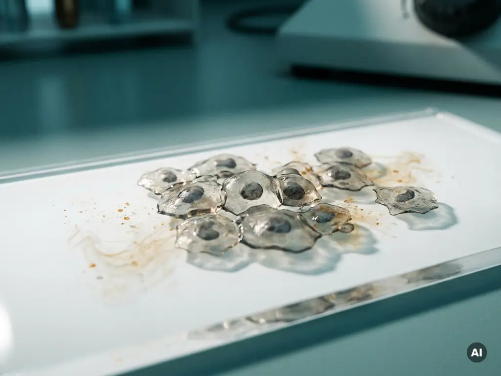
Intrinsic drug resistance and acquired drug resistance are two distinct mechanisms by which cancer cells evade therapeutic agents. Intrinsic resistance is present before any treatment begins, rooted in pre-existing genetic or cellular characteristics. Acquired resistance develops during or after treatment, as cancer cells adapt under selective pressure from the therapy itself.
Understanding the difference between these two resistance types is central to oncology research, because each demands a different experimental approach to study and, ultimately, a different strategy to address. The sections below unpack the mechanisms, research methods, and scientific directions that define this field in 2026.
How do cancer cells become resistant to treatment?
Cancer cells become resistant to treatment through a combination of pre-existing cellular traits and adaptive responses triggered by therapeutic pressure. Whether resistance is present from the start or emerges over time, the underlying principle is the same: a subpopulation of cells survives treatment and proliferates, eventually dominating the tumor.
Resistance can operate through several overlapping mechanisms. These include alterations in the drug target itself, activation of alternative survival pathways, changes in how cells absorb or expel drug molecules, and shifts in the tumor microenvironment that reduce drug accessibility. In some cases, cancer cells also acquire the ability to evade programmed cell death, meaning even high drug concentrations fail to trigger apoptosis.
Researchers studying cancer drug resistance need experimental models that faithfully replicate these dynamics. Three-dimensional culture systems, patient-derived organoids, and co-culture platforms have become increasingly important tools in this context, as they capture aspects of tumor biology that standard two-dimensional cell line models can miss.
What causes intrinsic drug resistance in cancer?
Intrinsic drug resistance in cancer is caused by genetic, epigenetic, or phenotypic characteristics that exist within a tumor before any therapeutic agent is introduced. Because these traits are already present at baseline, the cancer does not respond to the drug from the very first exposure.
Several molecular mechanisms underlie intrinsic resistance:
- Mutations in drug targets: If the protein a drug is designed to inhibit carries a mutation that reduces drug binding affinity, the therapy may be ineffective from the outset.
- Overexpression of efflux pumps: Some cancer cells constitutively express membrane transporters that actively remove drug molecules before they can reach their intracellular targets.
- Tumor heterogeneity: Within a single tumor, distinct subpopulations may carry different genetic profiles. A resistant subclone present before treatment begins may be selected once sensitive cells are eliminated.
- Epigenetic silencing: Genes that would otherwise sensitize cells to a drug may be silenced through methylation or chromatin remodeling, reducing drug efficacy without any change in DNA sequence.
- Microenvironmental factors: Physical barriers such as dense extracellular matrix or hypoxic regions can limit drug penetration into the tumor mass.
From a research perspective, studying intrinsic resistance requires baseline characterization of tumor samples before any treatment is applied. Genomic sequencing, transcriptomic profiling, and proteomic analysis of untreated tumor material help researchers identify which features correlate with poor initial response.
What causes acquired drug resistance in cancer?
Acquired drug resistance in cancer develops after treatment has begun, as cancer cells that survive initial therapy undergo genetic or epigenetic changes that allow them to tolerate or neutralize the drug. This form of resistance is an evolutionary process driven by selective pressure.
The mechanisms behind acquired resistance often mirror those seen in intrinsic resistance, but they arise de novo during treatment rather than existing beforehand. Key drivers include:
- Secondary mutations in the drug target: A new mutation may emerge in the gene encoding the drug’s target, altering the binding site and reducing drug efficacy. This is well documented in targeted therapy research.
- Pathway bypass: Cancer cells may activate alternative signaling routes that circumvent the inhibited pathway, maintaining proliferative signaling despite effective drug binding at the original target.
- Phenotypic plasticity: Some cancer cells transition between states, for example from an epithelial to a mesenchymal phenotype, adopting characteristics associated with greater stress tolerance and reduced drug sensitivity.
- Clonal evolution: Under the selective pressure of treatment, resistant clones that were initially a minor fraction of the tumor expand and eventually dominate the cell population.
- Epigenetic reprogramming: Cells may undergo heritable changes in gene expression patterns that confer resistance without any underlying mutation, making this form of resistance potentially reversible under the right experimental conditions.
Longitudinal experimental models are particularly valuable for studying acquired resistance, as they allow researchers to track how cell populations evolve over repeated drug exposures and identify the molecular events that accompany the transition from sensitivity to resistance.
What’s the difference between intrinsic and acquired resistance in clinical outcomes?
From a research standpoint, the key distinction between intrinsic and acquired resistance lies in their timing and origin. Intrinsic resistance means a tumor fails to respond from the beginning of treatment, while acquired resistance means an initially responsive tumor later stops responding as resistant cell populations emerge and expand.
This distinction has significant implications for experimental design and the development of therapeutic strategies. Researchers investigating intrinsic resistance focus on identifying biomarkers present in pre-treatment samples that predict non-response. This typically involves comparing molecular profiles of tumors that did not respond with those that did, looking for consistent differentiating features.
Acquired resistance research, by contrast, requires paired sample analysis, comparing tumor material collected before treatment with material collected at the point of resistance. This approach allows researchers to pinpoint the molecular changes that occurred during treatment and contributed to the shift in drug sensitivity.
Understanding this distinction also shapes how combination therapy strategies are designed in research settings. Studies exploring intrinsic resistance often test whether adding a second agent at baseline can overcome the pre-existing barrier. Studies of acquired resistance frequently investigate whether rotating or combining agents can prevent or delay the emergence of resistant clones.
How do researchers detect and study drug resistance mechanisms?
Researchers detect and study drug resistance mechanisms using a range of molecular, cellular, and imaging-based approaches that together build a picture of how cancer cells survive drug exposure. No single method captures the full complexity of resistance, so most research programs integrate multiple complementary techniques.
Genomic and transcriptomic profiling
Whole-genome sequencing, targeted panel sequencing, and RNA sequencing allow researchers to identify mutations, copy number variations, and changes in gene expression that distinguish resistant cells from sensitive ones. Comparing pre- and post-treatment samples is a standard approach for mapping the molecular landscape of acquired resistance.
Functional and phenotypic assays
Cell viability assays, apoptosis measurements, and proliferation tracking under varying drug concentrations help quantify resistance at the functional level. High-content imaging platforms and live-cell analysis systems enable researchers to monitor how cell populations respond to drug treatment over time, capturing dynamic changes that endpoint assays may miss. Flow cytometry is frequently used to characterize subpopulations within heterogeneous cultures and to measure markers associated with resistance phenotypes.
Three-dimensional tumor models, including spheroids and organoids derived from patient material, are increasingly used because they replicate aspects of drug penetration and microenvironmental influence that flat cell cultures cannot reproduce. These models allow researchers to study resistance in a context that more closely reflects the architecture of a real tumor.
Can drug resistance in cancer be overcome or prevented?
Drug resistance in cancer cannot always be fully prevented, but research suggests that its onset may be delayed and its mechanisms may be exploitable once understood. Several scientific strategies are actively being investigated to address both intrinsic and acquired resistance.
Combination approaches are among the most studied. By targeting multiple pathways simultaneously, researchers aim to reduce the probability that any single resistant subclone can survive and expand. Sequential treatment strategies, in which different agents are applied in a defined order, are also being explored as a way to outpace the evolutionary dynamics of resistance.
Epigenetic resistance mechanisms are of particular interest because they are potentially reversible. Research into agents that modify chromatin structure or DNA methylation patterns may open avenues for resensitizing cells that have become resistant through epigenetic reprogramming rather than fixed genetic mutation.
Tumor heterogeneity remains one of the most significant challenges. Because a tumor contains many genetically distinct subpopulations, a therapy effective against the dominant clone may leave resistant minor clones free to expand. Single-cell sequencing and spatially resolved transcriptomics are providing researchers with tools to map this heterogeneity at unprecedented resolution, potentially informing more precisely targeted experimental strategies.
Organoid and co-culture platforms that incorporate immune cells and stromal components are also helping researchers investigate how the tumor microenvironment contributes to resistance and whether modulating it may restore drug sensitivity. This remains an active and rapidly developing area of research across Benelux academic and biotech institutions.
How BioSPX supports cancer drug resistance research
Studying drug resistance in cancer requires experimental platforms that can capture cellular heterogeneity, dynamic responses, and complex three-dimensional biology. BioSPX provides life science research teams across Belgium, the Netherlands, and Luxembourg with access to instruments suited to exactly these demands.
- Imaging and high-content analysis systems for monitoring cell populations and resistance phenotypes over time
- Multimode readers for quantifying viability, apoptosis, and functional assay readouts across drug concentration gradients
- Liquid handling solutions for reproducible, high-throughput drug treatment workflows
- Organoid and 3D culture-compatible platforms for more physiologically relevant resistance modeling
- Expert product specialists who advise on instrument selection, assay configuration, and experimental design
- Installation, training, and maintenance support to keep research workflows running with minimal disruption
Whether your team is characterizing baseline resistance markers or tracking the emergence of acquired resistance across treatment cycles, BioSPX can help you identify the right instrumentation for your research goals. Contact BioSPX to discuss your experimental setup with a specialist.
Frequently Asked Questions
What experimental model should I start with if I'm new to studying drug resistance in cancer?
If you’re just beginning to study cancer drug resistance, starting with established 2D cell line models is practical for building foundational assay skills, but plan to transition quickly to 3D spheroid or organoid systems for more physiologically meaningful data. Patient-derived organoids are particularly valuable because they preserve the genetic heterogeneity of the original tumor, giving you a more realistic picture of resistance dynamics. Pairing these models with a multimode reader for viability and apoptosis readouts will cover most early-stage experimental needs.
How do I distinguish between a cell population that is intrinsically resistant and one that has simply been enriched through clonal selection during treatment?
This is one of the more nuanced challenges in resistance research. The key is pre-treatment characterization: if resistant subclones can be detected in untreated samples through single-cell sequencing or flow cytometric sorting, that points toward intrinsic heterogeneity and clonal selection rather than de novo acquired resistance. Longitudinal sampling at multiple time points during treatment, combined with clonal tracking tools, helps you map whether resistant populations were pre-existing or emerged as new variants under selective pressure.
Can epigenetic resistance be reversed, and how would I test this experimentally?
Epigenetic resistance is considered potentially reversible because it involves heritable changes in gene expression patterns rather than fixed DNA mutations, making it an attractive experimental target. To test reversibility, researchers typically withdraw the therapeutic agent for a defined period and then re-expose cells to assess whether drug sensitivity is restored. Combining this approach with chromatin accessibility assays such as ATAC-seq or DNA methylation profiling before and after drug withdrawal can help pinpoint which epigenetic marks correlate with the resistance and recovery states.
What are the most common mistakes researchers make when designing acquired resistance studies?
One of the most frequent pitfalls is using a single, acute high-dose drug exposure rather than a gradual, chronic exposure protocol, which better mimics the selective pressure cancer cells experience clinically. Another common mistake is failing to collect and biobank pre-treatment samples, which makes it impossible to perform the paired comparisons essential for identifying what changed during treatment. Researchers also sometimes overlook the role of the culture microenvironment, studying resistance in standard 2D monocultures without accounting for how stromal or immune components influence drug sensitivity.
How does tumor heterogeneity complicate the interpretation of drug resistance assay results?
Tumor heterogeneity means that bulk assay readouts, such as a population-level IC50 value, can mask the behavior of distinct subpopulations within the same culture. A small resistant subclone may not significantly shift the bulk viability curve initially, yet it is precisely that subclone that will dominate after prolonged drug exposure. Incorporating single-cell analysis tools, high-content imaging to resolve subpopulation morphology, or flow cytometry-based sorting alongside bulk assays gives a more complete picture and reduces the risk of underestimating resistance potential.
Is it possible to model both intrinsic and acquired resistance in the same experimental workflow?
Yes, and doing so in a structured way is actually good experimental design. You can begin by characterizing your cell model at baseline to identify any pre-existing resistance markers, which addresses intrinsic resistance, and then subject the same model to repeated or escalating drug exposures over weeks to generate an acquired resistance phenotype. Comparing the molecular profiles at baseline, during treatment, and at the point of established resistance within a single longitudinal experiment allows you to distinguish what was already present from what emerged, providing a more complete mechanistic picture.
What role does the tumor microenvironment play in drug resistance, and how can it be incorporated into in vitro research models?
The tumor microenvironment, including cancer-associated fibroblasts, immune cells, and the extracellular matrix, can physically limit drug penetration and biochemically promote resistance through paracrine signaling that activates survival pathways in cancer cells. Incorporating these components into in vitro models requires co-culture platforms that support multiple cell types simultaneously, or the use of scaffold-based 3D systems that replicate matrix stiffness and architecture. Organoid models that include stromal or immune cell fractions are increasingly used for this purpose and represent one of the most active areas of development in resistance research instrumentation.
Related Articles
- Why is spatial context important for understanding molecular mechanisms?
- How can image segmentation improve quantitative spatial biology analysis?
- What are the key differences between spatial proteomics and spatial transcriptomics?
- What is the difference between apoptosis and necrosis in cancer cells?
- What is the difference between benign and malignant tumors?
This content was generated with the help of AI and it may contain mistakes