
 EN

Cell migration and cell invasion are related but distinct processes in cancer biology. Cell migration refers to the directed movement of cancer cells through open spaces or along surfaces, while cell invasion describes the active penetration of cells through physical barriers such as the extracellular matrix or basement membrane. The critical distinction lies in whether a cell is simply moving or actively degrading and remodeling the tissue around it. Understanding this difference is essential for researchers studying metastasis, and the sections below explore the mechanisms, measurement methods, and research implications of each process.
How do cancer cells physically move through tissue?
Cancer cells move through tissue by generating mechanical forces, reorganizing their cytoskeleton, and interacting with the surrounding extracellular matrix. This movement is rarely passive. Cells actively sense chemical gradients, mechanical cues, and contact signals from neighboring cells or matrix components, then respond by extending protrusions, contracting their body, and detaching from their trailing edge to advance in a given direction.
The cytoskeletal machinery driving this movement relies primarily on actin polymerization at the cell front and myosin-driven contraction at the rear. Adhesion molecules, particularly integrins, anchor the cell to matrix fibers and transmit the traction forces needed to pull the cell body forward. The stiffness and composition of the surrounding matrix strongly influence how efficiently cancer cells can move, which is why the tumor microenvironment plays such a prominent role in cancer cell motility research.
Cells can also move collectively, maintaining adhesive contacts with neighboring cells while advancing as a coordinated sheet or strand. This collective behavior is especially relevant in epithelial tumors, where groups of cells may invade surrounding tissue together rather than as isolated individuals.
What is the key difference between cell migration and cell invasion?
The key difference between cell migration and cell invasion is whether the cell must degrade a physical barrier to move. Cell migration describes movement through pre-existing open spaces, along surfaces, or within loose connective tissue. Cell invasion requires the active breakdown of structured barriers, such as the basement membrane or dense extracellular matrix, using proteolytic enzymes.
In practical terms, a migrating cancer cell exploits gaps and channels that already exist in the tissue. An invading cancer cell creates its own path by secreting matrix metalloproteinases and other proteases that dissolve the surrounding matrix. This enzymatic remodeling is what makes invasion the more aggressive and clinically significant behavior, as it allows cancer cells to breach anatomical compartments and enter the bloodstream or lymphatic system.
Because invasion requires an additional biochemical capability beyond motility alone, not all migratory cancer cells are invasive. A cell can be highly motile yet unable to penetrate a structured matrix if it lacks sufficient protease activity or the cytoskeletal organization required for force generation against a resistant barrier.
Which modes of invasion do cancer cells use?
Cancer cells use several distinct invasion strategies depending on their molecular profile, the physical properties of the surrounding tissue, and signals from the tumor microenvironment. Researchers have identified at least three major invasion modes that are relevant to tumor invasion mechanisms.
- Mesenchymal invasion: Cells adopt an elongated morphology, form actin-rich protrusions called invadopodia, and rely heavily on matrix metalloproteinases to degrade the extracellular matrix ahead of them. This mode is associated with epithelial-to-mesenchymal transition.
- Amoeboid invasion: Cells move with a rounded, deformable shape and squeeze through matrix pores rather than degrading them. This mode is faster, less dependent on proteases, and can emerge when protease activity is pharmacologically blocked, representing a potential resistance mechanism.
- Collective invasion: Groups of cells advance together, with leader cells at the front performing matrix degradation while follower cells benefit from the path created. This mode is common in carcinomas and may be more representative of how many tumors invade surrounding stroma in vivo.
The ability of cancer cells to switch between these modes, a phenomenon sometimes called invasion plasticity, poses a significant challenge for researchers developing anti-invasive therapeutic strategies. Studying which modes predominate in a given cancer type and under what conditions cells transition between them is an active area of metastasis research.
How are migration and invasion measured in the lab?
Migration and invasion are measured using distinct assay formats that reflect the physical differences between the two processes. Choosing the right assay is fundamental to generating reproducible, interpretable data in cancer cell motility studies.
Migration assays
The scratch assay, also called the wound healing assay, is one of the most widely used migration assays. A gap is created in a confluent cell monolayer, and closure of that gap is monitored over time, typically using live-cell imaging. This format is straightforward and compatible with standard laboratory equipment, though it measures collective migration rather than individual cell movement.
Transwell migration assays place cells on an uncoated porous membrane. Cells that migrate through the pores to the underside of the membrane are counted after a defined period. This format allows researchers to study migration toward a chemoattractant gradient placed in the lower chamber.
 Real-time cell analysis can provide a quantitative way to monitor migration kinetics. The xCELLigence RTCA DP uses label-free impedance measurements in an electronically integrated Boyden chamber to continuously track cell movement, allowing researchers to compare migratory responses over time.
Invasion assays
Invasion assays use the same Transwell format but coat the membrane with a layer of reconstituted basement membrane matrix. Because cells must enzymatically degrade this barrier before passing through, only genuinely invasive cells are detected. The matrix coating is the defining feature that separates an invasion assay from a migration assay in practice.
Three-dimensional invasion assays embed cells in or on top of hydrogel matrices that more closely mimic tissue architecture. These formats allow researchers to observe invasion morphology, track individual cell trajectories, and measure invasion depth, providing richer spatial data than traditional membrane-based approaches.
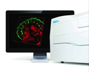 High-content imaging can provide complementary spatial information by allowing researchers to visualize how cells move and interact with their surrounding environment. The Cytation C10 can support migration and invasion studies in 2D and 3D models, helping researchers characterize changes in cell morphology, position, and interactions within an extracellular matrix.
Why does the migration-invasion distinction matter for drug discovery?
The distinction between migration and invasion matters for drug discovery because a compound that blocks cell motility may have no effect on invasive capacity, and vice versa. Researchers developing anti-metastatic strategies need to know precisely which step in the metastatic cascade their candidate compound is targeting.
If a drug inhibits actin dynamics and reduces migration speed but does not affect protease secretion, cancer cells may still be capable of penetrating the basement membrane even though they move more slowly. Conversely, a protease inhibitor may block matrix degradation without affecting the underlying motility machinery, leaving cells capable of amoeboid invasion through existing matrix pores. Testing compounds in both migration and invasion assays, rather than one alone, provides a more complete picture of their anti-metastatic potential.
This distinction also matters when interpreting published data. A study reporting reduced migration in response to a compound does not automatically support a conclusion about reduced invasiveness. Researchers reviewing the literature or designing their own experiments benefit from scrutinizing which assay format was used and what biological process it actually measures.
When should researchers study migration, invasion, or both?
Researchers should study migration alone when the scientific question concerns cell motility, chemotaxis, or wound healing responses that do not involve barrier penetration. Invasion assays become necessary when the research question relates to basement membrane crossing, matrix remodeling, or behavior that is directly relevant to in vivo tumor spreading. Studying both processes together is advisable when the goal is to characterize a cancer cell line comprehensively or evaluate a compound’s effect across multiple steps of metastasis.
The choice also depends on the cancer type and the biological context being modeled. Carcinomas that originate in epithelial compartments must cross a basement membrane to become invasive, making invasion assays highly relevant. For cancers arising in connective tissue, where no basement membrane barrier is present, migration assays may be more informative for the early stages of spread.
- Define the specific biological question and the step of metastasis under investigation.
- Select the assay format that best models the relevant barrier or tissue environment.
- Include both migration and invasion readouts if the study involves compound screening or phenotypic characterization.
- Validate findings in a three-dimensional or more physiologically relevant model where resources allow.
For researchers in the Benelux working across academic, pharmaceutical, or biotech settings, aligning assay selection with the biological question from the outset saves time and ensures that results are interpretable and reproducible across experimental replicates.
How BioSPX supports migration and invasion research
BioSPX helps research teams in the Benelux select, implement, and optimize the laboratory instruments needed to study cancer cell migration and invasion with confidence. Whether a lab is setting up a live-cell imaging workflow for scratch assays, introducing automated liquid handling to improve consistency in Transwell-based invasion assays, or moving toward three-dimensional culture models, BioSPX’s product specialists can advise on the most appropriate instrumentation for each experimental context.
- Expert guidance on imaging, liquid handling, and cell culture instruments suited to migration and invasion workflows
- Support for experimental setup, including assay optimization and instrument configuration
- Installation, user training, and rapid maintenance response through a dedicated service team
- ISO 9001-certified quality standards, with offices in the Netherlands and Belgium
If you are designing or scaling a migration or invasion research program and want to discuss which instruments best fit your workflow, contact BioSPX to speak with a product specialist.
Frequently Asked Questions
Can the same cancer cell line switch between migration and invasion depending on experimental conditions?
Yes, and this plasticity is one of the most important variables to account for when designing experiments. Factors such as matrix stiffness, oxygen availability, growth factor concentrations, and even cell density can shift a cell line’s dominant behavior. This is why characterizing a cell line under your specific culture conditions, rather than relying solely on published phenotypic descriptions, is strongly recommended before committing to a single assay format.
What are the most common mistakes researchers make when setting up a Transwell invasion assay for the first time?
The most frequent errors involve inconsistent matrix coating thickness, seeding too many or too few cells, and failing to optimize the chemoattractant concentration in the lower chamber. Using an expired or improperly thawed basement membrane matrix is another common source of variability that can produce misleading results. Running a migration control alongside every invasion assay, using uncoated membranes in parallel, helps confirm that any reduction in cell numbers reflects impaired invasion rather than reduced viability or motility.
How do I know whether a 2D assay is sufficient or whether I need to move to a 3D invasion model?
2D assays are appropriate for initial screening, relative comparisons between conditions, or studies where throughput is a priority. A 3D model becomes necessary when you need spatial information about invasion depth, when you are modeling a tissue architecture that cannot be represented by a flat membrane, or when 2D results are inconsistent with in vivo observations. As a practical rule, if your findings will inform in vivo or clinical decisions, validating key results in a 3D format adds meaningful confidence to your conclusions.
Are matrix metalloproteinase inhibitors still a viable therapeutic strategy given the amoeboid escape mechanism?
MMP inhibitors as standalone therapies have largely underperformed in clinical trials, partly because cancer cells can switch to protease-independent amoeboid invasion when protease activity is blocked. Current research is exploring combination strategies that simultaneously target both mesenchymal and amoeboid invasion modes, as well as upstream regulators of invasion plasticity such as Rho GTPase signaling. Understanding which invasion mode predominates in a specific tumor type before selecting a therapeutic target is now considered essential for designing more effective anti-invasive strategies.
How should I normalize my migration and invasion data to account for differences in cell proliferation?
Proliferation can inflate migration and invasion readouts, particularly in longer assays, making it difficult to determine whether increased cell numbers reflect genuine motility or simply faster division. Including a proliferation control, such as a parallel well treated with a mitotic inhibitor like mitomycin C, allows you to isolate motility-specific effects. Alternatively, shortening the assay duration or selecting a time window where proliferation contributes minimally to gap closure or membrane crossing can reduce this confounding variable.
What imaging capabilities are needed to get quantitative data from live-cell scratch assays?
At minimum, you need a microscope with environmental control (temperature, CO₂, and humidity) and automated stage positioning to capture the same field of view repeatedly over time. Software capable of automated gap area quantification significantly reduces manual analysis time and inter-user variability. For more advanced studies, systems with phase-contrast or brightfield imaging combined with single-cell tracking algorithms allow you to extract individual cell velocity and directionality data alongside bulk wound closure measurements.
How can I make my migration and invasion assay results more reproducible across different experimental runs?
Reproducibility in these assays depends heavily on standardizing every upstream variable: passage number, seeding density, serum starvation duration, and the lot number of any matrix material used. Automating liquid handling steps, particularly matrix coating and cell seeding, substantially reduces technician-to-technician variability. Establishing a detailed standard operating procedure for each assay format and validating it with a well-characterized positive control cell line before beginning experimental work is the most reliable way to ensure consistent, comparable results over time.
Related Articles
- How does FLIM-FRET preserve spatial information that bulk assays lose?
- How can researchers measure real-time changes in cancer cell morphology?
- What can continuous cell monitoring reveal that endpoint assays cannot?
- How do clinical trials work in oncology?
- What is the difference between curative and palliative cancer treatment?
This content was generated with the help of AI and it may contain mistakes