
 EN
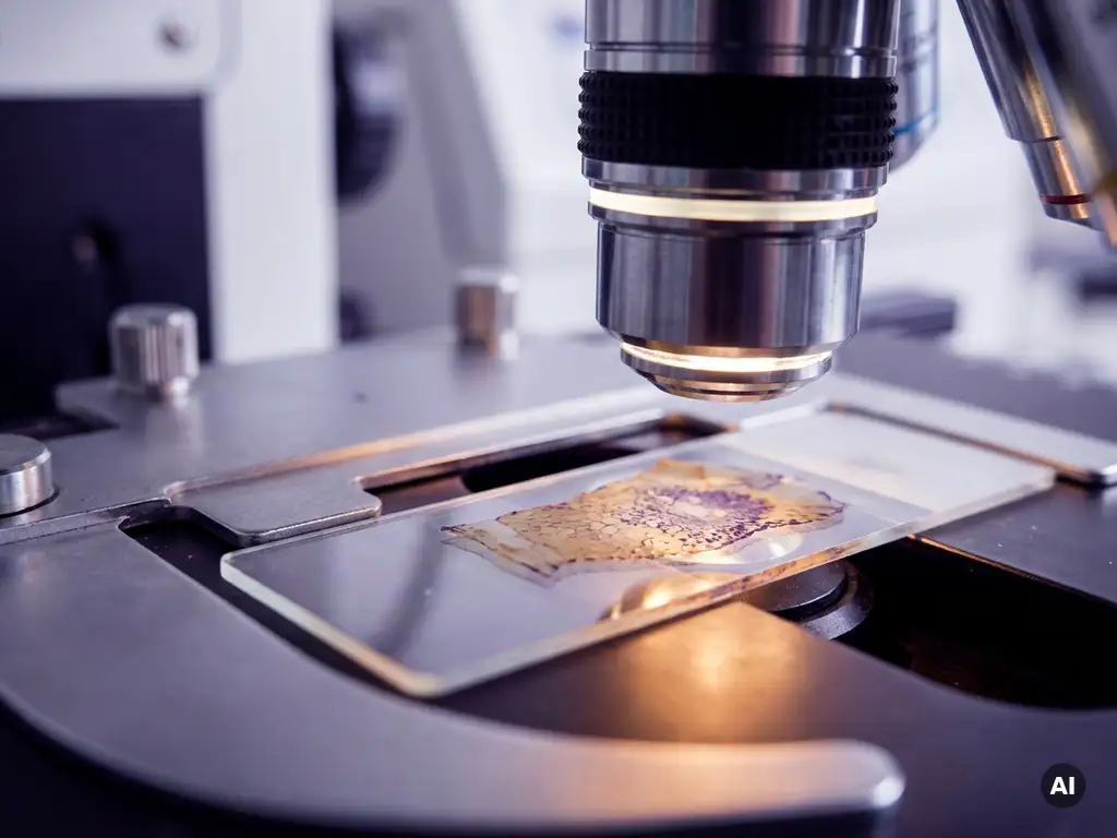
In situ analysis preserves cellular and tissue information by examining biological material within its native structural environment, rather than breaking it apart for analysis. This approach keeps cells spatially organized within the tissue, allowing researchers to study gene expression, protein distribution, and cell morphology in the context of surrounding architecture. The sections below explore how this works, which techniques are involved, and when in situ approaches offer a clear methodological advantage.
What types of biological information does in situ analysis capture?
In situ analysis captures a wide range of biological information that would otherwise be lost when tissue is dissociated or homogenized. It simultaneously preserves spatial context, cell morphology, molecular identity, and the structural relationships between cell populations within intact tissue or cellular environments.
More specifically, in situ techniques can reveal where a gene is expressed within a tissue section, which cell types carry a particular protein, how cell populations are distributed relative to one another, and how tissue architecture relates to molecular activity. This combination of spatial and molecular data is what makes in situ analysis particularly valuable for understanding complex biological systems.
- Gene expression patterns: which genes are active in specific cell types or tissue regions
- Protein localization: where proteins are found within cells and tissues, including subcellular compartments
- Cell morphology: the physical shape, size, and structural features of individual cells
- Tissue architecture: the spatial organization of cell layers, niches, and structural boundaries
- Cell-to-cell relationships: proximity, interaction zones, and microenvironmental composition
For researchers in the Benelux working in fields such as developmental biology, oncology research, or immunology, this multi-dimensional readout is often essential for generating biologically meaningful data.
How does in situ analysis keep cells and tissue intact during processing?
In situ analysis keeps cells and tissue intact by using fixation, embedding, and sectioning protocols that stabilize biological structures before any molecular analysis is performed. Rather than lysing or dissociating cells, the tissue is chemically preserved so that spatial relationships and cellular architecture remain undisturbed throughout the analytical workflow.
Fixation with agents such as formaldehyde crosslinks proteins and nucleic acids in place, preventing degradation and maintaining structural integrity. The tissue is then typically embedded in a support medium and cut into thin sections that can be mounted on slides for imaging or molecular detection. These sections retain the full spatial context of the original sample.
Subsequent steps, such as probe hybridization, antibody staining, or fluorescent labeling, are performed directly on these sections. Because the cells remain in their original positions throughout, the resulting data reflects genuine biological organization rather than an averaged or reconstructed signal from a mixed cell population.
What’s the difference between in situ analysis and dissociation-based methods?
The key difference between in situ analysis and dissociation-based methods is spatial context. In situ analysis studies cells and molecules within intact tissue, preserving their location and relationships. Dissociation-based methods, such as bulk RNA sequencing or single-cell sequencing after tissue digestion, break the tissue apart and lose all positional information in the process.
Dissociation-based approaches offer their own advantages. They can profile large numbers of cells rapidly, detect rare cell types with high sensitivity, and generate genome-wide molecular data. However, the process of enzymatic or mechanical dissociation introduces several limitations.
Dissociation can alter gene expression profiles as cells respond to stress during processing. It also removes any information about where a cell was located within the tissue, which microenvironment it occupied, and which neighboring cells it was interacting with. For research questions that depend on understanding tissue organization, these are significant losses.
In situ analysis sacrifices some throughput and molecular breadth in exchange for spatial fidelity. The two approaches are increasingly used together, with dissociation-based data providing molecular depth and in situ data providing the spatial map to interpret it within a tissue context.
Which in situ techniques are used for gene expression and protein detection?
The most widely used in situ techniques for gene expression detection are in situ hybridization methods, while immunohistochemistry and immunofluorescence are the primary approaches for protein detection. Both categories can be applied to fixed tissue sections and allow molecular signals to be visualized and mapped within the preserved tissue architecture.
In situ hybridization works by introducing labeled nucleic acid probes that bind to complementary RNA sequences within cells. Fluorescence-based variants, often referred to as fluorescence in situ hybridization or FISH, allow multiple targets to be detected simultaneously using spectrally distinct labels. More recent multiplexed in situ hybridization platforms can profile dozens to hundreds of RNA targets within a single tissue section, making them powerful tools for spatial transcriptomics workflows.
For protein detection, immunofluorescence uses fluorescently labeled antibodies that bind to specific protein targets, enabling their visualization under a fluorescence microscope. Multiplexed immunofluorescence approaches allow researchers to detect many proteins simultaneously by cycling through antibody panels or using spectrally resolved imaging systems.
Spatial biology platforms that combine high-plex in situ molecular detection with high-resolution imaging are an area of active development, and researchers in the Benelux are increasingly incorporating these approaches into studies of tissue organization, tumor microenvironments, and developmental processes.
When should researchers choose in situ analysis over other approaches?
Researchers should choose in situ analysis when the spatial location of a molecular signal is as important as the signal itself. If the research question requires knowing where a gene is expressed within a tissue, how protein distribution relates to cell morphology, or how different cell types are organized relative to one another, in situ methods are the appropriate choice.
There are several situations where in situ analysis offers a clear methodological advantage:
- When studying heterogeneous tissues where different regions have distinct biological identities, such as tumor cores versus invasion fronts
- When investigating cell-to-cell communication that depends on physical proximity
- When sample availability is limited and tissue architecture must be preserved for multiple analyses
- When validating findings from bulk or single-cell sequencing by confirming spatial patterns in tissue
- When studying rare cell populations that are defined by their location within a specific tissue niche
Dissociation-based methods remain preferable when broad molecular profiling across many cells is the priority, or when spatial context is not relevant to the research question. Many modern research workflows use both approaches in combination to generate a more complete biological picture.
How is imaging technology advancing in situ spatial resolution?
Imaging technology is advancing in situ spatial resolution by enabling molecular detection at subcellular and even nanoscale resolution, while also increasing the number of targets that can be visualized simultaneously. These advances are expanding what researchers can learn from a single tissue section without sacrificing spatial fidelity.
Super-resolution fluorescence microscopy techniques have pushed imaging resolution well beyond the diffraction limit of conventional light microscopy, making it possible to resolve individual RNA molecules or protein complexes within cells. When combined with in situ hybridization or immunofluorescence, these techniques allow researchers to study molecular organization at a level of detail that was previously inaccessible.
Multiplexed imaging platforms are another area of rapid development. By cycling through multiple rounds of staining and imaging, or by using spectrally distinct probes, researchers can now detect tens to hundreds of molecular targets within a single tissue section. This transforms in situ imaging from a targeted validation tool into a discovery platform capable of generating rich, spatially resolved molecular atlases.
Computational image analysis is also advancing in parallel, with machine learning approaches helping researchers extract quantitative data from complex, high-dimensional imaging datasets. For cellular analysis workflows in Benelux research institutions, these combined hardware and software developments are making spatial biology an increasingly practical and informative methodology.
How BioSPX supports in situ analysis and spatial biology research
BioSPX helps researchers in the Benelux access the imaging and cellular analysis instrumentation needed to implement in situ techniques effectively. Whether a laboratory is establishing a spatial biology workflow for the first time or expanding an existing setup, BioSPX offers expert guidance and a curated portfolio of solutions suited to these applications.
- Imaging and microscopy systems for fluorescence-based in situ detection
- Multimode readers and high-content imaging platforms for quantitative spatial analysis
- Liquid handling solutions to support reproducible staining and hybridization workflows
- Product specialists who advise on instrument selection, experimental setup, and system configuration
- Installation, training, and maintenance support from a dedicated service team
BioSPX serves academic research laboratories, biotech companies, pharmaceutical R&D teams, and other life science organizations across Belgium, the Netherlands, and Luxembourg. To discuss which imaging or cellular analysis solutions best fit your in situ research needs, contact the BioSPX team directly.
Frequently Asked Questions
How do I know if my tissue samples are compatible with in situ analysis workflows?
Most tissue types are compatible with in situ analysis, but sample quality is critical. Fresh-frozen and formalin-fixed paraffin-embedded (FFPE) tissues are the most commonly used formats, with FFPE being particularly practical due to its long-term stability and widespread availability in biobanks. The key factors to assess are fixation quality, RNA integrity (for hybridization-based methods), and section thickness, as poor fixation or over-processing can compromise probe binding efficiency and antibody performance. If you are working with archived FFPE samples, it is worth running a small optimization experiment before committing to a full study.
What are the most common mistakes researchers make when setting up an in situ hybridization experiment?
One of the most frequent pitfalls is insufficient optimization of probe hybridization conditions, particularly temperature and wash stringency, which can lead to high background signal or weak target detection. Inadequate tissue permeabilization is another common issue, as probes need to penetrate the cell membrane to reach their RNA targets. Researchers also often underestimate the importance of RNase-free working conditions when performing RNA-based in situ hybridization, since RNA degradation during sample preparation will significantly reduce signal quality. Starting with a well-validated positive control probe and a known-positive tissue section is strongly recommended before moving to novel targets.
Can in situ analysis be combined with single-cell sequencing data, and if so, how?
Yes, integrating in situ spatial data with single-cell RNA sequencing (scRNA-seq) data is one of the most powerful strategies in modern spatial biology. A common approach is to use scRNA-seq data to define cell type identities and gene expression signatures, then use in situ hybridization or spatial transcriptomics to map those cell types back onto the tissue section. Computational tools such as Seurat, Squidpy, and RCTD are specifically designed to facilitate this kind of data integration. The result is a spatially resolved cell atlas that combines the molecular depth of sequencing with the positional accuracy of in situ imaging.
How many targets can multiplexed in situ platforms realistically detect in a single tissue section?
The number of detectable targets varies significantly depending on the platform and detection chemistry used. Standard multiplexed immunofluorescence panels typically resolve 7 to 10 protein targets per section, while cycling-based approaches such as CODEX or Opal can extend this to 40 or more markers. For RNA detection, highly multiplexed in situ hybridization platforms like RNAscope HiPlex or commercial spatial transcriptomics solutions can profile anywhere from a few dozen to several hundred RNA targets simultaneously. When planning a multiplexed experiment, it is important to consider not only the number of targets but also panel design, spectral overlap, and the imaging system’s capacity to resolve and quantify each channel accurately.
What imaging system specifications should I prioritize when setting up an in situ fluorescence workflow?
The most important specifications to consider are fluorescence channel availability, spatial resolution, and throughput capacity. For multiplexed workflows, you will need a system with enough spectrally distinct channels to accommodate your probe or antibody panel without significant bleed-through. If subcellular localization is part of your research question, a confocal or super-resolution system will provide the optical sectioning and resolution needed to distinguish compartments reliably. For high-content or large-area tissue scanning, automated slide scanning systems with tiling capabilities are preferable. Consulting with an instrumentation specialist before purchasing is advisable, as the right configuration depends heavily on your specific assay format and throughput requirements.
Is in situ analysis suitable for clinical or translational research settings, or is it primarily a discovery tool?
In situ analysis is well-established in both discovery and translational research contexts. Immunohistochemistry, one of the most widely used in situ protein detection methods, has been a standard tool in clinical pathology for decades. In translational oncology, in situ techniques are routinely used to characterize tumor microenvironments, assess biomarker expression, and validate findings from genomic studies in patient-derived tissue. Multiplexed spatial biology approaches are increasingly being adopted in clinical trial settings to identify predictive biomarkers and understand treatment response at the tissue level. The key consideration for translational applications is ensuring that the workflow is reproducible, quantitative, and compatible with the sample types available in a clinical cohort.
How long does a typical in situ analysis experiment take from sample preparation to final data?
The timeline depends on the technique and the complexity of the experiment, but most in situ workflows span two to five days from sample preparation through to image acquisition. FFPE sample preparation, including sectioning and deparaffinization, typically takes half a day to a full day, followed by antigen retrieval or protease treatment. Hybridization or antibody incubation steps often run overnight, and imaging time scales with the number of targets, tissue area, and resolution required. Computational analysis and image quantification add additional time, particularly for high-plex or large-scale datasets. Automated liquid handling and slide scanning systems can significantly reduce hands-on time and improve reproducibility across larger experiments.
Related Articles
- How can researchers combine tissue mapping with molecular interaction measurements?
- What can spatial data analysis reveal about cellular heterogeneity?
- What is clonal evolution and how does it shape tumor heterogeneity?
- What are the most common types of cancer treated in oncology?
- How do clinical trials work in oncology?
This content was generated with the help of AI and it may contain mistakes