
 EN
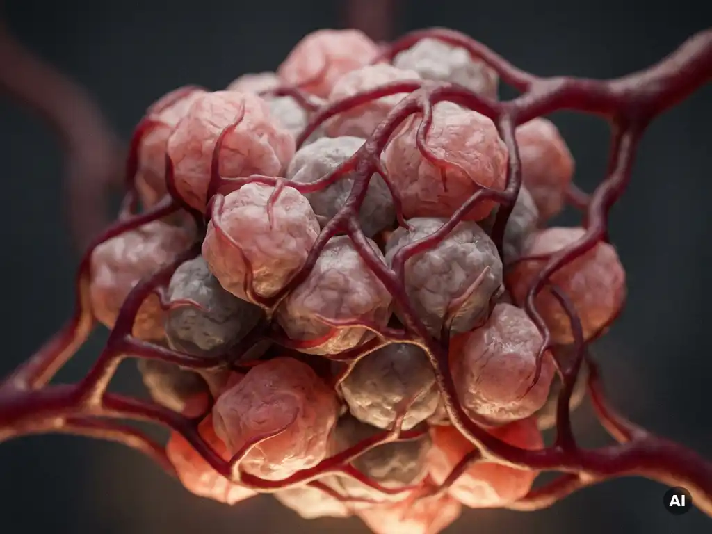
Angiogenesis supports tumor growth by supplying developing cancer tissue with oxygen, nutrients, and a route for waste removal, enabling tumors to expand beyond the size limits imposed by passive diffusion. Without new blood vessel formation, solid tumors typically stall at a few millimeters in diameter. Once a tumor triggers angiogenesis, it gains the vascular infrastructure needed to grow aggressively and, eventually, to spread. The sections below unpack the mechanisms, structural features, and research approaches that make tumor angiogenesis one of the most intensively studied areas in cancer biology.
What triggers angiogenesis in a tumor microenvironment?
Angiogenesis in a tumor is triggered when rapidly proliferating cancer cells outpace the local oxygen supply, creating a hypoxic microenvironment. This oxygen deficit activates hypoxia-inducible factors (HIFs), transcriptional regulators that switch on a cascade of pro-angiogenic signaling. The tumor microenvironment then shifts from an angiogenic-suppressed state to an angiogenic-permissive one, a transition researchers call the angiogenic switch.
Several cell types within the tumor microenvironment contribute to this switch. Cancer-associated fibroblasts, tumor-associated macrophages, and infiltrating immune cells all release pro-angiogenic mediators in response to hypoxic and inflammatory signals. At the same time, the balance between pro-angiogenic factors and natural inhibitors such as thrombospondin-1 tips decisively toward vessel formation. Understanding which signals dominate in different tumor types is an active area of investigation, particularly for researchers exploring how the composition of the tumor microenvironment influences disease progression.
How does VEGF drive new blood vessel formation in tumors?
Vascular endothelial growth factor (VEGF) is the primary molecular driver of tumor angiogenesis. When secreted by hypoxic tumor cells, VEGF binds to receptors on nearby endothelial cells, stimulating their proliferation, migration, and organization into new vessel sprouts. This process, known as sprouting angiogenesis, rapidly extends the tumor’s vascular network toward nutrient-rich regions.
VEGF does more than initiate vessel growth. It increases vascular permeability, allowing plasma proteins to leak into the surrounding tissue and create a provisional matrix that supports further endothelial migration. VEGF also recruits bone-marrow-derived progenitor cells that contribute to nascent vessel walls. Because VEGF signaling is central to the angiogenesis cancer mechanism, it has become one of the most studied molecular targets in cancer research. Laboratories investigating this pathway often use receptor-binding assays, reporter gene systems, and endothelial cell sprouting models to dissect how VEGF isoforms and their receptors interact under different microenvironmental conditions.
What makes tumor blood vessels structurally different from normal vessels?
Tumor blood vessels are structurally abnormal compared to the organized, hierarchical vascular networks found in healthy tissue. They are irregular in shape, poorly branched, and often lack the complete pericyte coverage that stabilizes normal capillaries. Their basement membranes are thickened and disorganized, and the endothelial cells lining them are loosely connected, resulting in a leaky and dysfunctional vascular bed.
These structural differences have direct functional consequences that researchers study closely:
- Irregular vessel geometry creates uneven blood flow, leaving some tumor regions persistently hypoxic despite apparent vascularization
- High interstitial fluid pressure, caused by leaky vessels, can impair the delivery of experimental agents to tumor tissue in preclinical models
- Absent or incomplete pericyte coverage makes tumor vessels more susceptible to collapse and remodeling
- Abnormal endothelial junctions allow large molecules to pass through vessel walls more readily than in normal tissue
These features are not merely incidental. They shape the entire biology of the tumor microenvironment and influence how researchers design in vitro and in vivo models intended to replicate the conditions cancer cells actually experience.
How do tumor blood vessels enable metastasis?
Tumor blood vessels enable metastasis by providing cancer cells with a physical route into the systemic circulation. The leaky, poorly formed endothelial junctions characteristic of cancer blood vessels make it easier for tumor cells to intravasate, entering the bloodstream or lymphatic system and traveling to distant sites. This process is a critical step in the metastatic cascade that researchers are working to understand at the molecular level.
Beyond simple physical access, the abnormal tumor vasculature actively promotes metastatic behavior. Hypoxic regions generated by irregular blood flow upregulate genes associated with epithelial-to-mesenchymal transition, a process through which cancer cells acquire a more migratory and invasive phenotype. Endothelial cells in tumor vessels also express adhesion molecules that facilitate the attachment and extravasation of circulating tumor cells at secondary sites. Studying these interactions in controlled laboratory settings, using microfluidic vessel-on-a-chip models or transwell invasion assays, allows researchers to isolate specific steps in the metastatic process and test how different microenvironmental conditions influence them.
What are the main anti-angiogenic strategies used in cancer research?
The main anti-angiogenic strategies investigated in cancer research target the molecular signals, cellular processes, or structural components that tumors exploit to build new blood vessels. Researchers approach this from several angles, and the field continues to evolve as new mechanisms are identified.
- VEGF pathway inhibition: Blocking VEGF or its receptors disrupts the primary signaling axis driving tumor vessel formation. This remains one of the most studied approaches in preclinical angiogenesis research.
- Receptor tyrosine kinase targeting: Small molecules that inhibit multiple pro-angiogenic receptor tyrosine kinases simultaneously are widely used in experimental tumor models to assess how broadly suppressing angiogenic signaling affects vascular growth.
- Vascular normalization: Rather than eliminating tumor vessels entirely, some research strategies aim to structurally normalize them, improving blood flow and reducing hypoxia. This approach is being explored for its potential to improve the delivery of other experimental agents in preclinical settings.
- Targeting pericyte recruitment: Interfering with the signals that recruit pericytes to stabilize nascent vessels may destabilize the tumor vasculature and is an emerging area of investigation.
- Endogenous inhibitors: Naturally occurring anti-angiogenic proteins such as endostatin and angiostatin are studied as potential research tools for understanding how the body normally suppresses vessel growth.
Each of these strategies generates distinct experimental questions about tumor vascular biology, and researchers in the Benelux are actively contributing to this field through work conducted in academic institutions, biotech organizations, and pharmaceutical R&D environments.
How is angiogenesis studied in the laboratory?
Angiogenesis is studied in the laboratory using a range of in vitro, ex vivo, and in vivo models that capture different aspects of the vessel formation process. Each model system offers specific readouts and is chosen based on the experimental question being addressed, whether that is initial endothelial cell activation, three-dimensional vessel sprouting, or the interaction between tumor cells and vascular structures.
Common laboratory approaches include endothelial tube formation assays on extracellular matrix substrates, aortic ring assays that preserve the multicellular architecture of vessel sprouting, and spheroid-based sprouting models that allow three-dimensional analysis. More advanced platforms such as microfluidic chips and organoid-based vascular models are gaining traction in research settings because they more closely replicate the spatial and mechanical conditions of the tumor microenvironment.
Imaging plays a central role in angiogenesis research. Fluorescence microscopy, live-cell imaging, and high-content imaging systems allow researchers to quantify vessel length, branching density, and endothelial cell behavior in real time or at defined time points. Automated image analysis tools improve the reproducibility and throughput of these measurements, which is particularly valuable when screening multiple experimental conditions or evaluating the effects of different pro- and anti-angiogenic compounds in parallel.
How BioSPX supports angiogenesis research
Studying tumor angiogenesis demands instruments that can reliably capture the complexity of vascular biology, from single-cell imaging to high-throughput functional assays. BioSPX provides life science laboratories in the Benelux with the instrumentation and expert support needed to build and maintain these workflows.
- Multimode readers and imaging systems suited to tube formation, sprouting, and permeability assays
- High-content imaging platforms for automated, reproducible quantification of vascular network parameters
- Liquid handling solutions that improve consistency across multi-condition angiogenesis experiments
- Cell culture automation tools, including solutions for organoid and 3D model maintenance
- Installation, user training, and ongoing technical support from product specialists with deep application knowledge
Whether your laboratory is establishing a new angiogenesis model or scaling up an existing workflow, BioSPX’s team can advise on the most suitable instrument configuration for your research needs. Contact BioSPX to discuss your laboratory requirements with a specialist.
Frequently Asked Questions
What is the difference between sprouting angiogenesis and intussusceptive angiogenesis in tumors?
Sprouting angiogenesis, the most widely studied form, involves endothelial cells migrating and proliferating outward from existing vessels to form new sprouts guided by VEGF gradients. Intussusceptive angiogenesis, by contrast, expands the vascular network by splitting existing vessels from within through the insertion of tissue pillars, without requiring significant endothelial cell proliferation. Tumors can exploit both mechanisms, and some evidence suggests that intussusception becomes more prominent after anti-angiogenic treatment, which is why researchers are increasingly designing assays that can detect and distinguish between the two modes of vessel growth.
How do I choose the right in vitro angiogenesis model for my research question?
The choice of model depends on which step of the angiogenic process you want to interrogate. Endothelial tube formation assays on Matrigel are well suited for rapid screening of pro- or anti-angiogenic compounds, while aortic ring assays and spheroid sprouting models better preserve the multicellular context of vessel formation and are preferable when studying stromal interactions or three-dimensional sprouting behavior. If your research involves the tumor microenvironment or metastatic intravasation, microfluidic vessel-on-a-chip platforms offer greater physiological relevance, though they require more specialized equipment and expertise to implement reliably.
Why do some tumors stop responding to anti-angiogenic therapies over time?
Resistance to anti-angiogenic therapy typically arises through one or more adaptive mechanisms: tumors can upregulate alternative pro-angiogenic signaling pathways that bypass VEGF blockade, recruit additional bone-marrow-derived cells to sustain vessel formation, or shift toward vessel co-option, in which cancer cells exploit pre-existing host vasculature rather than forming new vessels. Persistent hypoxia caused by vascular pruning can paradoxically select for more aggressive, treatment-resistant tumor cell populations. Understanding these resistance mechanisms is an active area of preclinical research, and models that allow long-term monitoring of vascular remodeling under treatment conditions are particularly valuable for studying them.
What role do pericytes play in tumor angiogenesis, and why does their absence matter experimentally?
Pericytes are mural cells that wrap around endothelial cells to provide structural support, regulate vessel tone, and reinforce the blood-brain barrier and other specialized vascular beds. In tumor vasculature, pericyte coverage is often incomplete or functionally abnormal, which contributes to vessel leakiness, irregular perfusion, and instability. Experimentally, this matters because pericyte deficiency affects how faithfully an in vitro or in vivo model replicates true tumor vascular function, and it is also a potential therapeutic target: disrupting pericyte recruitment signals can destabilize nascent tumor vessels, while promoting pericyte coverage is central to vascular normalization strategies.
How can high-content imaging improve the reproducibility of angiogenesis assays?
High-content imaging platforms automate image acquisition and analysis across large numbers of wells or conditions, removing the subjectivity and fatigue-related variability associated with manual microscopy. For angiogenesis assays specifically, automated quantification of parameters such as total tube length, branching points, mesh area, and sprout number produces consistent, objective data that is directly comparable across experiments and between laboratories. This is particularly valuable when running dose-response experiments with multiple anti-angiogenic compounds in parallel, as it significantly increases throughput without sacrificing the spatial detail needed to characterize vascular network morphology.
What are the most common mistakes researchers make when setting up a tube formation assay?
The most frequent pitfalls include working with Matrigel or similar extracellular matrix substrates at room temperature, which causes premature polymerization and uneven gel distribution across the well. Using endothelial cells at too high a passage number is another common issue, as late-passage cells lose their angiogenic responsiveness and produce inconsistent or absent tube networks. Timing is also critical: tubes typically reach their optimal morphology between four and eight hours after seeding, and imaging too early or too late will misrepresent the extent of network formation. Standardizing cell passage number, substrate handling temperature, and imaging time points before beginning a multi-condition experiment will significantly improve data quality and reproducibility.
Are organoid-based vascular models ready for routine use in angiogenesis research, or are they still primarily experimental?
Vascular organoid models, including self-organizing endothelial-pericyte organoids and tumor organoids with embedded vascular components, have advanced considerably and are now used in a growing number of research laboratories, though they remain more technically demanding than conventional two-dimensional or aortic ring assays. Their primary advantage is the ability to study vessel formation within a three-dimensional, multicellular architecture that more closely resembles the in vivo tumor microenvironment, making them well suited for mechanistic studies and for testing how vascular structure responds to stromal or immune cell interactions. For laboratories considering adoption, starting with established protocols and investing in appropriate imaging and culture automation infrastructure will help bridge the gap between proof-of-concept use and routine experimental application.
Related Articles
- How does FLIM-FRET preserve spatial information that bulk assays lose?
- What should researchers consider when analyzing FFPE tissue samples?
- What is the tumor microenvironment and why does it matter in research?
- How do genetic mutations contribute to cancer development?
- How do oncologists decide which cancer treatment to recommend?
This content was generated with the help of AI and it may contain mistakes