
 EN
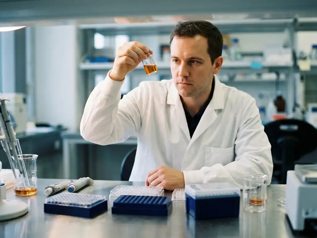
Clinical trials in oncology are structured research studies that test whether new cancer therapies, diagnostic approaches, or treatment strategies are safe and effective in human participants. They follow a defined sequence of phases, each designed to answer specific scientific questions before a therapy can advance further. The sections below unpack the key questions researchers, scientists, and procurement professionals in the Benelux region encounter when working with or around oncology clinical research.
What are the different phases of an oncology clinical trial?
Oncology clinical trials are organized into four main phases, each serving a distinct scientific purpose. Phase I focuses on safety and dosing, Phase II on early signals of activity, Phase III on comparative effectiveness at scale, and Phase IV on long-term monitoring after broader use. Together, these phases form a structured pathway for evaluating new cancer-related interventions.
Each phase builds on the evidence generated in the previous one:
- Phase I: A small group of participants receives the investigational therapy to assess tolerability, identify safe dosage ranges, and observe how the body processes the compound. The primary goal is safety, not therapeutic outcome.
- Phase II: A larger cohort is enrolled to gather preliminary data on biological activity and to further characterize the safety profile. Researchers begin to examine whether the therapy produces measurable effects relevant to the cancer type under investigation.
- Phase III: The investigational therapy is compared against the current standard of care or a placebo in a large, often multicenter trial. This phase generates the core evidence base that informs regulatory review.
- Phase IV: Conducted after a therapy has received regulatory authorization, this phase monitors real-world performance, rare adverse events, and long-term outcomes across broader patient populations.
In oncology specifically, trial design has grown more sophisticated. Adaptive trial designs, basket trials (which group participants by molecular target rather than tumor type), and umbrella trials are increasingly used to accelerate the evaluation of targeted therapies and immunotherapies.
Who decides which patients are eligible to join a cancer trial?
Eligibility for a cancer clinical trial is determined by a set of predefined inclusion and exclusion criteria established by the trial’s scientific and medical team. These criteria are based on factors such as cancer type, disease stage, prior treatment history, biomarker status, and general health indicators. Their purpose is to ensure participant safety and scientific validity.
Inclusion criteria define the characteristics a participant must have, such as a confirmed diagnosis of a specific cancer subtype or expression of a particular molecular marker. Exclusion criteria identify conditions or circumstances that would disqualify participation, such as certain comorbidities or prior exposure to related therapies.
Biomarker-driven eligibility has become especially important in modern oncology trials. As precision oncology advances, many trials require molecular profiling of tumor tissue before enrollment. This is where laboratory instrumentation plays a direct role: genomic sequencing platforms, flow cytometry systems, and imaging tools are routinely used to confirm whether a candidate meets the biological criteria for a given trial.
What is randomization and blinding in oncology trials?
Randomization is the process of assigning trial participants to different treatment groups by chance rather than by choice, while blinding prevents participants and sometimes researchers from knowing which group a participant belongs to. Together, these methods reduce bias and increase the reliability of trial results.
In a randomized controlled trial, participants are allocated to either the investigational arm or the control arm (which may receive standard therapy or a placebo) through a random process. This prevents systematic differences between groups that could distort the findings.
Blinding adds another layer of scientific rigor. In a single-blind trial, participants do not know their group assignment. In a double-blind trial, neither participants nor the investigators assessing outcomes are aware of the assignment. Double-blind randomized controlled trials are considered the methodological gold standard in clinical research, including oncology, because they minimize the influence of expectation and observer bias on the data collected.
How are new cancer therapies evaluated for safety and effectiveness?
New cancer therapies are evaluated through a combination of preclinical laboratory studies, phased clinical trials, and predefined endpoints that measure specific biological or clinical outcomes. Safety is assessed continuously throughout the trial process, while effectiveness is measured against endpoints such as tumor response, disease progression markers, or survival metrics defined at the trial’s outset.
Before a therapy reaches human trials, it undergoes extensive laboratory and animal-based research to establish a basic safety profile and identify a plausible mechanism of action. Once a trial begins, independent data monitoring committees review accumulating safety data at regular intervals.
Effectiveness in oncology trials is typically assessed through:
- Objective response rate: The proportion of participants whose tumors show measurable reduction according to standardized imaging criteria.
- Progression-free survival: The length of time during and after treatment that a participant lives without disease progression.
- Overall survival: The length of time from enrollment that participants remain alive, considered the most definitive endpoint.
- Biomarker endpoints: Molecular or cellular indicators measured in blood, tissue, or other samples that reflect the biological activity of the therapy.
The choice of endpoint depends on the therapy’s mechanism, the cancer type, and the phase of the trial. Early-phase oncology trials frequently rely on biomarker and imaging endpoints, which is where laboratory instrumentation becomes central to the research workflow.
What’s the difference between investigator-initiated and industry-sponsored oncology trials?
Investigator-initiated trials are designed, conducted, and sponsored by academic researchers or clinical institutions, while industry-sponsored trials are funded and managed by pharmaceutical or biotechnology companies. The key distinction lies in who controls the scientific question, the protocol, and the data.
Investigator-initiated trials often explore novel scientific hypotheses, repurposed compounds, or combinations of existing therapies that may not yet have commercial interest. They are typically funded through academic grants or institutional budgets and are common in university hospitals and research centers across the Benelux region.
Industry-sponsored trials, by contrast, are designed to generate the evidence package needed to support regulatory review of a proprietary compound. These trials tend to be larger, more resource-intensive, and governed by the sponsor’s regulatory strategy. They often involve multiple sites across countries and require standardized laboratory protocols and data collection systems.
Both types of trial rely heavily on laboratory infrastructure. Investigator-initiated trials in particular depend on in-house analytical and imaging capabilities, making instrument quality and reproducibility critical to the integrity of the research.
What role does laboratory instrumentation play in clinical oncology research?
Laboratory instrumentation is foundational to clinical oncology research. It enables the biological measurements, sample analyses, and data generation that underpin every phase of a cancer clinical trial. From cell-based assays in early discovery to biomarker quantification in Phase III studies, instruments such as multimode readers, imaging systems, liquid handling platforms, and cell culture automation tools are integral to reproducible, high-quality oncology research.
In translational oncology research, which bridges laboratory findings and clinical application, instruments must deliver consistent, validated performance. Multimode plate readers are used to quantify cellular responses to investigational compounds. Imaging and microscopy systems support morphological and functional analysis of tumor models, including organoid-based platforms that are increasingly used as more physiologically relevant preclinical tools. Liquid handling systems reduce manual variability in sample preparation, which is particularly important when working with limited or precious clinical samples.
Cell culture automation is another area of growing relevance. As organoid and 3D tumor model research expands in oncology, maintaining consistent culture conditions across experiments becomes both labor-intensive and scientifically critical. Automated cell culture systems can help research teams maintain reproducibility across large experimental sets, supporting the quality standards that clinical-grade oncology research demands.
How BioSPX supports clinical oncology research in the Benelux
For research teams in Belgium, the Netherlands, and Luxembourg working within oncology clinical research environments, access to reliable, well-supported instrumentation is a practical necessity. BioSPX provides life science laboratory instruments and expert guidance specifically tailored to the needs of academic, biotech, pharmaceutical, and clinical research organizations across the Benelux region.
BioSPX’s portfolio and support services relevant to clinical oncology research include:
- Multimode readers and imaging systems for biomarker quantification and cellular analysis
- Liquid handling solutions that reduce manual variability in sample preparation workflows
- Cell culture automation tools, including systems designed to support organoid and 3D tumor model research
- Installation, user training, and rapid maintenance response through a dedicated service department
- Expert product specialists who advise on instrument configuration and experimental setup
Whether you are equipping a new oncology research laboratory or upgrading an existing workflow, the team at BioSPX can help you identify the right instruments for your scientific goals. Contact BioSPX to discuss your research requirements with a specialist.
Frequently Asked Questions
How long does a typical oncology clinical trial take from Phase I to regulatory approval?
The full journey from Phase I through regulatory approval typically spans 10 to 15 years, though this varies considerably depending on the therapy type, cancer indication, and trial outcomes. Oncology has seen some acceleration in recent years through adaptive trial designs, accelerated regulatory pathways (such as EMA’s PRIME scheme), and rolling submissions that allow review of completed data packages before the full dossier is finalized. Breakthrough designations and conditional marketing authorizations can also shorten timelines for therapies addressing unmet medical needs.
What are the most common reasons an oncology clinical trial fails or is discontinued?
The most frequent reasons include insufficient efficacy signals at Phase II, unexpected toxicity findings, poor patient enrollment or retention, and protocol deviations that compromise data integrity. In oncology specifically, a mismatch between the patient population enrolled and the therapy’s actual mechanism of action is a recurring challenge — one that better biomarker-driven patient selection at the design stage can help mitigate. Inadequate preclinical validation of endpoints and biomarkers is another upstream factor that contributes to late-stage failures.
How does molecular profiling of tumor tissue work in practice, and what instrumentation is involved?
Molecular profiling typically involves extracting nucleic acids or proteins from tumor biopsy material and analyzing them for specific mutations, gene expression patterns, or protein markers relevant to the trial’s eligibility criteria or endpoints. Next-generation sequencing (NGS) platforms, flow cytometry systems, immunohistochemistry imaging systems, and digital pathology tools are commonly used depending on the biomarker type and required sensitivity. Liquid biopsy approaches — analyzing circulating tumor DNA from blood samples — are also increasingly used, requiring highly sensitive detection platforms and rigorous sample preparation workflows to avoid pre-analytical variability.
What should a research team consider when selecting laboratory instruments for an oncology trial environment?
Key considerations include whether the instrument can be validated or qualified to the standards required for the trial (GLP, GCP, or ISO compliance depending on the context), the reproducibility and sensitivity of measurements across runs and operators, and the availability of service and calibration support — particularly important in regulated research environments. For teams in the Benelux region, local service infrastructure and response time are practical factors that directly affect research continuity. It is also worth evaluating whether the instrument’s data output is compatible with the trial’s data management and analysis systems from the outset.
Can organoid and 3D tumor models be used as part of a clinical trial workflow, or are they limited to preclinical research?
Currently, organoid and 3D tumor models are primarily used in the preclinical and translational phases of oncology research — for example, to model patient-specific drug responses or to validate biomarker hypotheses before clinical testing. However, patient-derived organoids (PDOs) are increasingly being explored as companion research tools alongside clinical trials, where tumor material from enrolled patients is cultured to study ex vivo drug sensitivity in parallel with in-trial outcomes. This is an active area of methodological development, and the reproducibility of organoid culture — supported by automated cell culture systems — is critical to generating data that can meaningfully complement clinical findings.
How do investigator-initiated trials in the Benelux region typically access the instrumentation they need?
Academic and clinical research teams in Belgium, the Netherlands, and Luxembourg generally source instrumentation through a combination of institutional procurement, grant-funded equipment budgets, and specialist life science distributors with regional expertise. Because investigator-initiated trials often operate with tighter budgets than industry-sponsored studies, instrument reliability, total cost of ownership, and access to local technical support are especially important selection criteria. Working with a regional specialist like BioSPX — rather than sourcing internationally without local service coverage — can significantly reduce downtime risk and ensure that instrument performance is maintained to the standard the research requires.
What is the difference between a primary endpoint and a secondary endpoint in an oncology trial, and why does it matter?
The primary endpoint is the main outcome measure that the trial is statistically powered and designed to evaluate — it is the result on which the trial’s success or failure is formally judged, and it directly informs regulatory submissions. Secondary endpoints capture additional outcomes of scientific or clinical interest, such as quality of life measures, duration of response, or exploratory biomarker findings, but the trial is not sized to definitively prove these. The distinction matters because claims made about a therapy’s effectiveness — including those reviewed by regulators — must be grounded in the pre-specified primary endpoint, not post-hoc analysis of secondary or exploratory data.
Related Articles
- How does FRET measure molecular proximity below the resolution limit of conventional microscopy?
- How can spatially resolved functional data help validate cellular hypotheses?
- What can spatial data analysis reveal about cellular heterogeneity?
- How can image segmentation improve quantitative spatial biology analysis?
- What causes genomic instability in cancer cells?
This content was generated with the help of AI and it may contain mistakes