
 EN
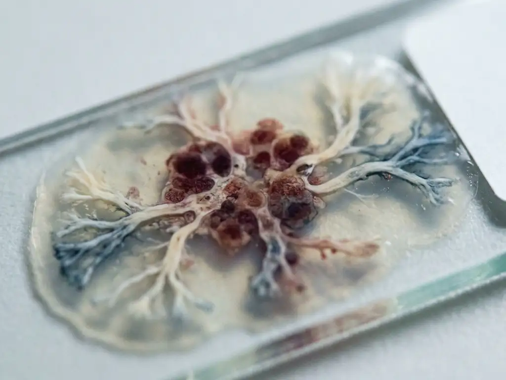
A primary cancer is the original tumor that develops in the tissue or organ where cancer first begins. A secondary cancer is a tumor that forms when cancer cells break away from the primary tumor and travel through the bloodstream or lymphatic system to establish new growths elsewhere in the body. The two tumors are biologically related: secondary cancer carries the same cell type as the primary, even when it appears in a completely different organ. The sections below explore how this process unfolds, where secondary tumors tend to appear, and what researchers are investigating to better understand and address metastatic disease.
How does a primary cancer become a secondary cancer?
A primary cancer becomes a secondary cancer through a process called metastasis. Cancer cells from the primary tumor acquire the ability to invade surrounding tissue, enter the bloodstream or lymphatic vessels, survive in circulation, and then exit into a distant tissue where they begin to proliferate. This multi-step biological cascade is what distinguishes a localized primary tumor from systemic, metastatic disease.
The process is not random. Metastasis depends on a series of molecular events, including changes in cell adhesion proteins, the secretion of enzymes that degrade surrounding tissue, and the ability of cancer cells to evade immune surveillance while in transit. Research into the mechanisms driving each of these steps is an active area of oncology and cancer biology.
Once cancer cells reach a distant site, they must adapt to a new microenvironment. Many circulating tumor cells do not survive this process. Those that do may remain dormant for months or years before forming a detectable secondary tumor, which is one reason why secondary cancers can appear long after a primary tumor has been identified and addressed.
Where do secondary cancers most commonly spread?
Secondary cancers most commonly develop in the liver, lungs, bones, and brain. These organs receive large volumes of blood flow, which increases the likelihood of circulating tumor cells arriving and establishing themselves. The lymph nodes are also frequent sites of secondary spread, particularly in the early stages of metastasis.
The specific pattern of spread is strongly influenced by the origin of the primary tumor. Some well-documented patterns include:
- Breast cancer frequently spreads to bone, lung, liver, and brain
- Colorectal cancer most commonly metastasizes to the liver and lungs
- Prostate cancer shows a strong tendency to spread to bone
- Lung cancer can spread to the brain, adrenal glands, and liver
- Melanoma is known for spreading widely, including to the brain and gastrointestinal tract
This organ-specific tropism is thought to reflect molecular compatibility between cancer cells and the target tissue environment, a concept sometimes described as the “seed and soil” hypothesis in cancer biology.
Are primary and secondary cancers treated the same way?
Primary and secondary cancers are not treated the same way. While treatment strategies share some common tools, secondary cancer is generally approached differently because the cancer cell type of the secondary tumor reflects the primary origin, not the organ where it has spread. A secondary liver tumor originating from colorectal cancer, for example, is investigated and managed as colorectal cancer, not as primary liver cancer.
This distinction has significant implications for research and drug development. Therapies are typically directed at the molecular characteristics of the original cancer cell type. Understanding whether a tumor is primary or secondary is therefore essential for selecting the most relevant research models and experimental approaches.
Secondary cancer also tends to indicate a more advanced stage of disease, which influences the range of interventions being studied. Researchers are investigating systemic approaches, targeted molecular therapies, and immunological strategies, all of which require accurate classification of the tumor’s origin.
What are the symptoms of secondary cancer?
The symptoms of secondary cancer depend on which organ or tissue is affected. Because secondary tumors can form in many different parts of the body, the clinical signs vary widely. There is no single symptom pattern that applies to all secondary cancers.
Common symptom patterns associated with specific sites of secondary spread include:
- Bone metastases: Persistent bone pain, increased risk of fractures, and sometimes elevated calcium levels in the blood
- Liver metastases: Abdominal discomfort, jaundice, loss of appetite, and fatigue
- Lung metastases: Persistent cough, shortness of breath, and chest discomfort
- Brain metastases: Headaches, cognitive changes, visual disturbances, and neurological symptoms
In a research context, understanding the symptom profile associated with secondary tumors at specific sites helps investigators design studies that use appropriate models and endpoints. Imaging systems and cellular analysis tools play an important role in detecting and characterizing secondary tumor growth in preclinical and translational research settings.
Can secondary cancer be cured?
Secondary cancer is generally considered more difficult to resolve than primary cancer, and in many cases it is investigated as a chronic, systemic condition rather than a localized one. Whether secondary cancer can be fully addressed depends on a range of factors, including the type of primary cancer, the number and location of secondary tumors, and the biological characteristics of the cancer cells involved.
Research is ongoing into approaches that may contribute to better long-term outcomes in metastatic disease. These include targeted therapies directed at specific molecular drivers, immunological approaches that aim to enhance the body’s recognition of cancer cells, and combination strategies that address both the primary and secondary tumor environments simultaneously.
In some cases, particularly when secondary spread is limited to a single site, interventional research approaches are being investigated as potentially curative strategies. However, these scenarios remain the exception rather than the rule, and the field continues to explore what biological conditions make such outcomes possible.
What is the difference between recurrent cancer and secondary cancer?
Recurrent cancer is cancer that returns to the same site as the original primary tumor after a period of remission. Secondary cancer refers specifically to new tumor growth at a different location in the body, caused by the spread of cells from the primary tumor. The key distinction is location: recurrence is local or regional, while secondary cancer is distant and metastatic.
Both recurrent and secondary cancers involve the same cell type as the original primary tumor, which differentiates them from a completely new, unrelated primary cancer that may develop independently in a person who has previously had cancer.
From a research perspective, recurrence and secondary spread are studied through different biological lenses. Recurrence often reflects the survival of residual cancer cells that were not eliminated, while secondary spread involves the additional biological steps of invasion, circulation, and colonization at a distant site. Understanding these mechanisms separately is important for developing more precise research models and experimental frameworks.
How BioSPX supports cancer biology research in the Benelux
Studying the mechanisms behind primary and secondary cancer requires precise, reliable instrumentation. In the Benelux region, research teams investigating cancer metastasis, tumor biology, and cellular behavior rely on high-quality laboratory tools to generate reproducible, meaningful data.
BioSPX supports life science researchers across Belgium, the Netherlands, and Luxembourg with an extensive portfolio of instruments suited to cancer biology workflows. Relevant solutions for this field include:
- Imaging and microscopy systems for visualizing tumor cell behavior, invasion assays, and metastatic spread in model systems
- Multimode readers for cell viability, proliferation, and functional assays used in cancer research
- Liquid handling solutions that increase throughput and reproducibility in high-content screening workflows
- Automation platforms such as cell culture systems that reduce variability in long-term cancer cell maintenance
BioSPX also provides expert guidance from product specialists who advise on experimental setup, system configuration, and instrument selection. Installation, training, and responsive maintenance support are included as part of the service offering.
If you are setting up or expanding a cancer biology research program in the Benelux, contact BioSPX to discuss which instruments best fit your research needs.
Frequently Asked Questions
How do researchers detect secondary tumors in preclinical cancer models?
In preclinical research, secondary tumors are typically detected using a combination of in vivo imaging techniques (such as bioluminescence or fluorescence imaging), histological analysis of dissected tissues, and flow cytometry to identify circulating tumor cells. Advanced imaging systems allow researchers to monitor metastatic spread in real time within animal models, providing spatial and temporal data that is difficult to obtain through endpoint analysis alone. The choice of detection method depends on the model system used and the specific metastatic site being studied.
What is tumor dormancy, and why does it matter for understanding secondary cancer?
Tumor dormancy refers to the state in which disseminated cancer cells survive at a secondary site without actively proliferating, sometimes for months or even years. This is clinically significant because it helps explain why secondary tumors can emerge long after a primary cancer has been treated and the patient appears to be in remission. Researchers are actively investigating the molecular signals that keep dormant cells in a quiescent state and, critically, what triggers their reactivation — since disrupting that reactivation process could be a key strategy in preventing late-stage metastatic disease.
What is the 'seed and soil' hypothesis, and how does it guide metastasis research today?
The ‘seed and soil’ hypothesis, originally proposed by surgeon Stephen Paget in 1889, suggests that cancer cells (the ‘seeds’) do not spread randomly but instead preferentially colonize organs whose microenvironment (the ‘soil’) is biologically compatible with their survival and growth. Modern research has validated and greatly expanded this concept, identifying specific receptor-ligand interactions, chemokine gradients, and extracellular matrix properties that attract particular cancer cell types to specific organs. This framework continues to guide the development of targeted therapies aimed at disrupting the hospitable conditions that secondary sites provide to incoming tumor cells.
Can a person develop a completely new primary cancer that is unrelated to their original diagnosis?
Yes, a person who has had cancer can develop an entirely new, unrelated primary cancer — this is referred to as a subsequent primary cancer or second primary malignancy, and it is distinct from both recurrence and secondary (metastatic) cancer. Risk factors include genetic predisposition, prior exposure to radiation or chemotherapy, and lifestyle factors. Distinguishing a new primary cancer from a secondary tumor is critically important because the two require fundamentally different treatment approaches, and accurate classification relies on pathological analysis of the tumor’s cell type and molecular profile.
What laboratory assays are most commonly used to study cancer cell invasion and metastatic potential in vitro?
The most widely used in vitro assays for studying invasion and metastatic potential include the Transwell invasion assay (using Matrigel-coated membranes to mimic the extracellular matrix), wound-healing (scratch) assays to assess cell migration, and 3D spheroid invasion models that more closely replicate the tumor microenvironment than traditional 2D cultures. These assays are often paired with multimode plate readers and imaging systems to quantify cell movement, viability, and morphological changes. Selecting the right combination of assays and instrumentation is essential for generating reproducible data that accurately reflects metastatic behavior.
How important is reproducibility when studying metastasis, and what are common sources of variability in these experiments?
Reproducibility is especially critical in metastasis research because the biological processes involved — invasion, circulation, and colonization — are inherently variable and sensitive to subtle differences in experimental conditions. Common sources of variability include inconsistent cell culture conditions (passage number, confluency, media composition), operator-dependent pipetting errors in multi-step assays, and batch-to-batch differences in extracellular matrix reagents. Automated liquid handling systems and standardized cell culture platforms can significantly reduce these variables, improving the reliability and comparability of results across experiments and research groups.
At what stage of cancer does metastasis typically occur, and can it happen before a primary tumor is diagnosed?
Metastasis is most commonly associated with advanced-stage cancer, but the biological process of tumor cell dissemination can begin earlier than clinical detection suggests. In some cancer types, small numbers of tumor cells may enter the bloodstream at relatively early stages, potentially seeding distant sites while the primary tumor is still small or even before it is clinically detectable — a concept known as early dissemination. This is one reason why liquid biopsy research, which aims to detect circulating tumor cells or tumor DNA in blood samples, is a growing area of interest: it may offer a way to identify metastatic activity before secondary tumors become visible on imaging.
Related Articles
- How does in situ analysis preserve cellular and tissue information?
- What is the role of high-content imaging in cancer cell phenotyping?
- What is liquid biopsy and how is it used in oncology?
- What is the difference between benign and malignant tumors?
- How do oncologists monitor treatment progress?
This content was generated with the help of AI and it may contain mistakes