In our lab we primarily work with bacteria which typically lie in the range of 2-10 micrometer in size. Given the previous usage of the Lionheart FX with primarily eukaryotic cells which are much larger, we were keen to test the machine.
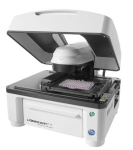 We started with imaging cell wall-deficient Streptomyces bacteria using both brightfield and fluorescence settings. Loss of the cell wall results in an unregulated form of cell division which is difficult to study and capture. The Lionheart imaging using timelapse control has been helpful for us in understanding the process of growth and division of these cells. The laser autofocus keeps cells in the right plane for imaging for extended periods of time. Thanks to the high-throughput imaging platform we have also imaged hyphal growth of various mutants of Streptomyces, fluorescent E. coli under different conditions and even entire colonies. This machine has provided us with a powerful operational range (from the colony to single cell level), the ability to image multiple conditions (96-well plate carriers) and and a super easy focus tracking (laser-trained autofocus). It is being used by multiple groups within the institute and helped initiate many exciting projects.
The software is easy to use and the employees at BioSPX have been very helpful with the different settings and our requirements. The response to any questions or issues is prompt with willingness to solve the problem.
Dr. Shraddha Shitut
Origins Center Fellow (post doc)
Institute of Biology (IBL) & Institute of Chemistry (LIC)
Leiden, The Netherlands