BioTek Cytation 5
Information
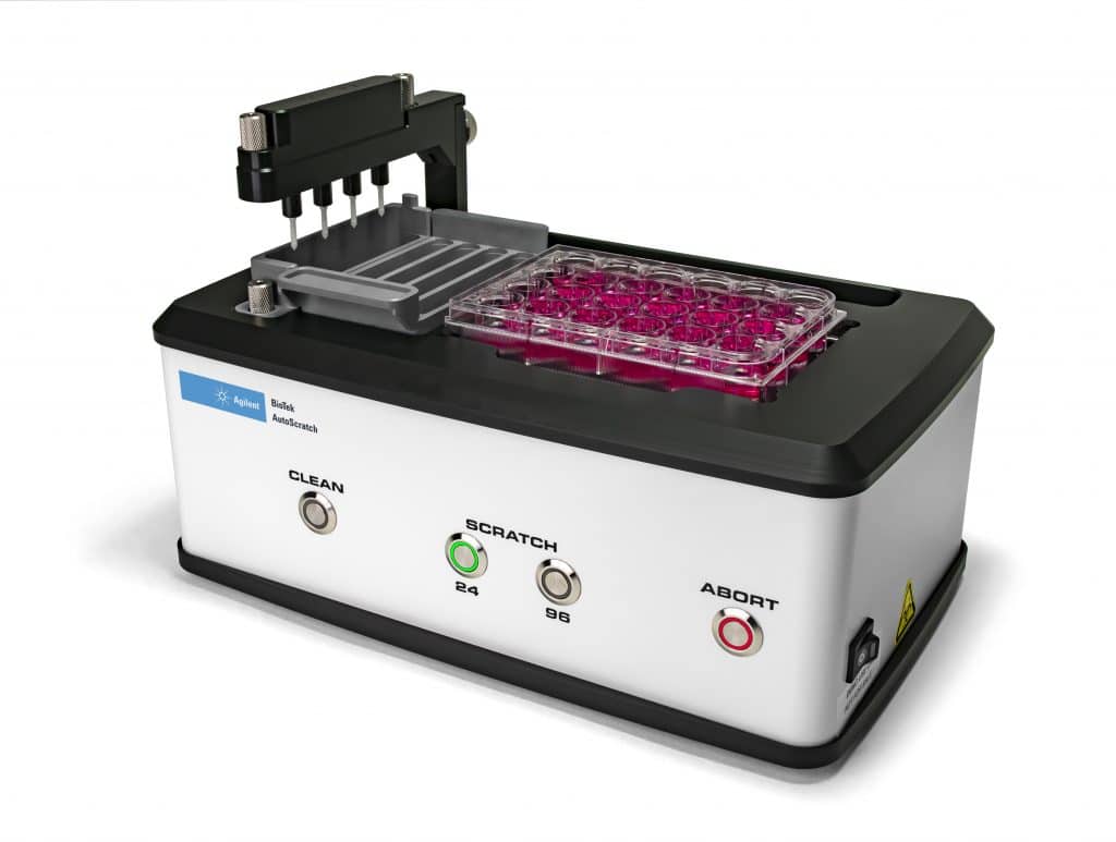
Cytation5 Cell Imaging Multi-Mode Reader combines automated digital widefield microscopy with conventional multi-mode microplate reading in a unique, patented design. With Cytation 5, you can capture rich phenotypic cellular information and well-based quantitative data in a single experiment.
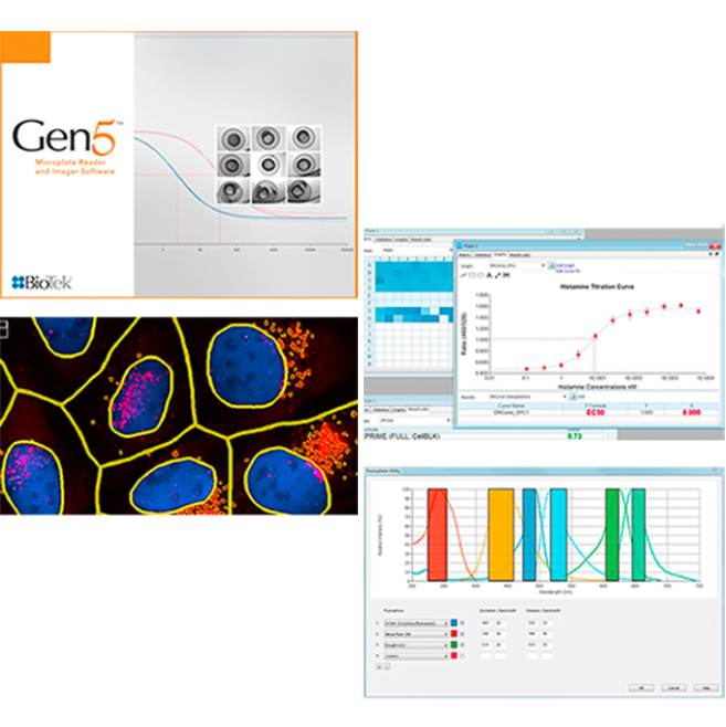
Cytation 5, along with Gen5 software, automates the workflow from image capture, processing, and analysis to create publication-ready images and data. Augmented Microscopy™ is the combination of all of these features in one compact system.
- Combined digital microscopy and multi-mode detection
- Patented Hybrid optics: sensitivity of filters, flexibility of monochromator
- Variable bandwidth monochromators optimize fluorophore detection
- 100 mW Alpha Laser: Speed and performance-based excitation for Alpha assays
- CO2/O2 control, incubation and shaking for live cell assays
- Label-free cell counting for cell proliferation studies
“The Cytation 5 helped us in the study of Macrophage metabolism in health and disease” – PhD Sanne Verberk, Department of Molecular Cell Biology and Immunology, Amsterdam UMC, VUmc
Read the testimonial here
-
Configurations
Imaging Plus Multi-Mode Configurations
Part # CYT5FV CYT5FW CYT5FAV CYTFAW CYT5MV CYT5MW CYT5MPV CYT5MPW CYT5MFV CYT5MFW CYT5MFAV CYT5MFAW Fluorescence imaging • • • • • • • • • • • • Brightfield imaging • • • • • • • • • • • • Color brightfield imaging • • • • • • • • • • • • Phase contrast imaging • • Wide FOV camera • • • • • • Filter-based fluorescence intensity • • • • • • • • Filter-based fluorescence polarization • • • • • • • • Filter-based time resolved fluorescence • • • • • • • • Filter-based luminescence • • • • • • • • Filter-based Alpha detection • • • • Monochromator-based UV-Vis absorbance • • • • • • • • Monochromator-based fluorescence intensity • • • • • • • • Monochromator-based time resolved fluorescence (secondary) • • • • • • • • Monochromator-based Luminescence • • • • • • • • Take3 Plate compatible • • • • • • • • Imaging Only and Multi-Mode Only Configurations
Part # CYT5V CYT5W CYT5PV CYT5PW CYT5MF CYT5MFA CYT5F CYT5FA CYT5M Fluorescence imaging • • • • Brightfield imaging • • • • Color brightfield imaging • • • • Phase contrast imaging • • Wide FOV camera • • Filter-based fluorescence intensity • • • • Filter-based fluorescence polarization • • • • Filter-based time resolved fluorescence • • • • Filter-based luminescence • • • • Filter-based Alpha detection • • Monochromator-based UV-Vis absorbance • • • Monochromator-based Fluorescence intensity • • • Monochromator-based Time resolved fluorescence (secondary) • • • Monochromator-based Luminescence • • • Take3 Plate compatible • • • -
Specifications
GENERAL Detection modes UV-Vis absorbance Fluorescence intensity Luminescence Fluorescence polarization Time-resolved fluorescence Alpha Read methods Endpoint, kinetic, spectral scanning, well area scanning Microplate types Monochromator: 6- to 384-well plates Filters: 6- to 1536-well plates Imaging: 6- to 1536-well plates Other labware supported Microscope slides, Petri and cell culture dishes, cell culture flasks (T25), counting chambers (hemocytometer) Take3 Micro-Volume Plates Temperature control 4-Zone incubation to 65 °C with Condensation Control +0.2 °C at 37 °C Shaking Linear, orbital, double orbital Software Gen5™ Microplate Reader and Imager Software Gen5 Image+ and Image Prime software available for full image analysis Gen5 Secure for 21 CFR Part 11 compliance (option) Automation BioStack and 3rd party automation compatible BioSpa 8 Automated Incubator compatible CO2 and O2 control (option) Range: 0 – 20% (CO2); 1 – 19% (O2) Models for both CO2 and O2 or CO2 only are available IMAGING SYSTEM Imaging mode Fluorescence Brightfield Color Brightfield Phase Contrast Imaging method Single color, multi-color, montage, time lapse, z-stacking Image processing Z-projection, digital phase contrast, stitching Camera 16-bit gray scale, Sony CCD Objective capacity 6 user-replaceable objectives Objectives available 1.25x, 2.5x (2.25x eff),2.5x (2.75x eff), 4x,10x, 20x, 40x, 60x Phase objectives available 4x, 10x, 20x, 40x Image filter cube capacity 4 user-replaceable fluorescence cubes plus brightfield channel Imaging filter cubes available DAPI, CFP, GFP, YFP, RFP, Texas Red, CY5, CY7,Acridine Orange (ACR OR), CFP-YFP FRET, propidium,Iodide, chlorophyll, phycoerythrin, CY5.5, TagBFP, Alexa568, Ex377 / Em647 Imaging LED cubes available 365 nm, 390 nm, 465 nm, 505 nm, 523 nm, 590 nm, 623 nm, 655 nm, 740 nm Automated functions Autofocus, auto LED intensity, auto exposure Autofocus method Image-based autofocus User-trained autofocus Laser autofocus (option) Positional controls Software control Joystick control (option) Image collection rate Image-based autofocus: 96 wells, 1 color (DAPI), 4x, 6 minutes 96 wells, 3 colors, 4x, 12 minutes Laser autofocus: 96 wells, 1 color (DAPI), 4x, <3 minutes 96 wells, 3 colors, 4x, <7 minutes, 30 seconds Burst Mode: 10 fps, single well, single color at <= 5Oms integration time Image Analysis Software option Gen5 Image+: Advanced image analysis Gen5 Image Prime: Advanced image analysis Gen5 Secure Image+: Advanced image analysis, 21 CFR Part 11 features FLUORESCENCE INTENSITY Light source Xenon flash Detector PMT for monochromator system PMT for filter system Wavelength selection Quad monochromators (top/bottom) Filters (top) Wavelength range Monochromators: 250 – 700 nm (850 nm option) Filters: 200 – 700 nm (850 nm option) Monochromator bandwidth Variable, from 9 nm to 50 nm in 1 nm increments Dynamic range 7 decades Sensitivity Filters: Fluorescein 0.25 pM (0.025 fmol/well, 384-well plate) Quad Monochromator: Fluorescein 2.5 pM (0.25 fmol/well, 384-well plate) – top Fluorescein 4 pM (0.4 fmol/well, 384-well plate) – bottom Reading speed (kinetic) 96 wells: 11 seconds 384 wells: 22 seconds LUMINESCENCE Wavelength range 300 – 700 nm Dynamic range >6 decades Sensitivity Monos: 20 amol ATP (flash) Filters: 10 amol ATP (flash), 100 amol (glow) FLUORESCENCE POLARIZATION Light source Xenon flash Detector PMT Wavelength selection Filters Wavelength range 280 – 700 nm (850 nm option) Sensitivity 1.2 mP standard deviation at 1 nM fluorescein TIME-RESOLVED FLUORESCENCE Light source Xenon flash Detector PMT Wavelength selection Quad monochromators (secondary mode) Filters (top) Wavelength range Filters: 200 – 700 nm (850 nm option) Sensitivity Filters: Europium 40 fM (4 amol/well, 384-well plate) Monos: Europium 1200 fM (120 amol/well, 384-well plate) ABSORBANCE Light source Xenon flash Detector photodiode Wavelength selection Monochromator Wavelength range 230 – 999 nm, 1 nm increment Monochromator bandwidth 4 nm (230 – 285 nm), 8 nm (>285 nm) Dynamic range 0 – 4.0 OD Resolution 0.0001 OD Pathlength correction yes Monochromator wavelength accuracy +2 nm Monochromator wavelength repeatability +0.2 nm OD accuracy <1% at 2.0 OD <3% at 3.0 OD OD linearity <1% from 0 to 3.0 OD OD repeatability <0.5% at 2.0 OD Stray light 0.03% at 230 nm Reading speed (kinetic) 96 wells: 11 seconds 384 wells: 22 seconds ALPHA DETECTION Light source 100 mW 680 nm laser Detector PMT Wavelength selection Filters (top) Sensitivity 100 amol LCK peptide (384-well low volume plate) REAGENT INJECTORS (OPTION) Supported detection modes All modes Number 2 syringe pumps Supported labware 6- to 384-well plates, Petri and cell culture dishes Dead volume 1.1 mL with back flush Dispense volume 5 – 1000 µL in 1 µL increments Plate geometry 6- to 384-well microplates, Petri dishes Dispense accuracy +1 µL or 2% Dispense precision <2% at 50 – 200 µL PHYSICAL CHARACTERISTICS Power 250 Watts max. Dimensions 20″ D x 16.5″ W x 17.5″ H (50.8 cm x 41.91 cm x 44.5 cm) Weight 80 lbs (36.3 Kg) REGULATORY Regulatory CE and TUV marked. RoHS Compliant. Models for In Vitro Diagnostic use are available. -
Peltier Cooling Module
The Peltier Cooling Module for Agilent’s Cytation™ 5 and Cytation 1 Cell Imaging Multi-Mode Readers helps maintain temperature stability inside the reading chamber, regardless of changes in ambient (room) temperature and heat produced by electro-mechanical operations within the reader.
The compact Peltier Cooling Module attaches easily to Cytation’s rear case. (See image)Instrument without Peltier Cooling Module:
The instrument’s interior temperature (red) follows ambient room temperature (green). It is also significantly warmer than ambient temperature because of heat produced by the instrument. This can result in assay data variation through the day, or drifts for kinetic assays run at ambient temperature Instrument with Peltier Cooling Module:
The instrument’s interior temperature (blue) is stabilized which results in more consistent assay data, and higher quality room temperature kinetic data Benefits of the Peltier Cooling Module for Cytation:
• Helps maintain the instrument’s internal ambient temperature stability, with less than 1 °C rise over external ambient, enabling more consistent results
• Quickly reduces internal temperature following incubated assays up to 3x faster, to allow both incubated and ambient assays to be run more efficiently in multi-user environments.Peltier Cooling Module is available for:
• Cytation Multi-Mode Reader
• Cytation 5 Cell Imaging Multi-Mode Reader
• Cytation 1 Cell Imaging Multi-Mode Reader -
Features
Combined digital microscopy and multi-mode detection
Cytation 5’s patented design incorporating automated digital microscopy with filter- and/or monochromator-based multi-mode detection is configurable – add modules as your research requirement change over time. Along with Gen5 software, Cytation captures both phenotypic and quantitative data within a single experiment.Patented Hybrid optics: sensitivity of filters, flexibility of monochromator
Cytation 5’s hybrid optical design offers filter-based fluorescence optics for high transmission and high sensitivity, while the available monochromator optics offers convenience and assay flexibility. Multiple fluorescence measurement modes include fluorescence intensity, time-resolved fluorescence and fluorescence polarizationVariable bandwidth monochromators optimize fluorophore detection
The bandwidths of the monochromators in Cytation 5’s fluorescence detection module are selectable from 9 nm to 50 nm, in 1 nm increments. Variable bandwidths allow you to optimize detection conditions for fluorophores that have unique excitation or emission parameters, for example, a narrow Stokes shift, to provide better performance than conventional monochromators.100 mW Alpha Laser: Speed and performance-based excitation for Alpha assays
Laser-based excitation offers the power required to get great sensitivity and fast reading speeds, which are critical for Alpha assays.CO2/O2 control, incubation and shaking for live cell assays
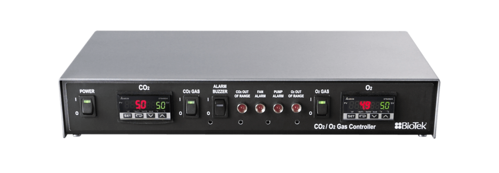
Cytation 5 is well suited for live cell assays, with temperature control to 65 °C, plus linear and orbital shaking. A gas controller module is available to help regulate CO2 and O2 levels in the system for longer-term live cell requirements. A dual reagent injector provides quick inject/image capability for observing fast reactionsLabel-free cell counting for cell proliferation studies
It’s fast and simple to count live cells using the available high contrast brightfield option. Higher contrast allows a direct and label-free method for object identification using Gen5 software to count cells and characterize cell proliferation in kinetic studies.
Brochures
Application notes
Please login to be able to see the application notes.