
 EN
Overview
Live cell imaging records living cells repeatedly to follow biological processes over time. Brightfield, phase contrast and fluorescence microscopy can reveal when responses begin, how quickly they develop and how individual cells differ. These measurements provide temporal information that a single endpoint image cannot recover.
Choosing a readout and acquisition schedule
The biological question determines the contrast method and sampling interval. Transmitted-light imaging can monitor morphology or surface coverage without a fluorescent label. Fluorescence enables molecularly specific readouts, such as reporter localisation or signals from validated functional probes. A readout that changes over minutes requires a different schedule from a proliferation assay lasting several days. Temporal sampling must be sufficient to capture the event while limiting unnecessary exposure.
Maintaining the cell environment
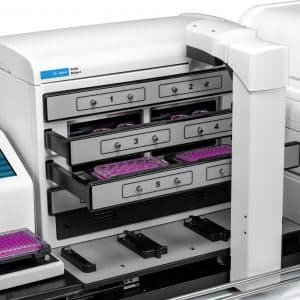
Temperature, medium pH, evaporation and gas conditions can change cell behaviour. For bicarbonate-buffered media, CO2 control is particularly relevant to pH stability. Oxygen control may be required for experiments investigating defined oxygen conditions. Suitable vessels, stable incubation and condensation management help maintain image quality and reproducibility. Agilent offers environmental controls on compatible Cytation and Lionheart configurations, and BioSpa 8 supports automated incubation between scheduled instrument operations. [1,2]
Managing photobleaching and phototoxicity
Photobleaching reduces fluorophore signal; phototoxicity changes the specimen biology. The two processes are related to illumination but are not interchangeable measures of damage. Experimental work has demonstrated that imaging conditions can alter biological development, so an attractive image alone does not establish that cells are unaffected. [3] Use the lowest exposure that supports the required measurement, minimise unnecessary channels and Z-sections, and compare with less frequently imaged or non-imaged controls where feasible.
Analysing a kinetic experiment
Maintain consistent acquisition and analysis settings, inspect focus and segmentation across time, and account for cell movement into or out of the field. Field selection should represent the culture rather than favour visually uniform regions. Technical fields and repeated frames are subsamples of an experiment; they do not replace independent biological replicates. Interpret intensity changes alongside cell number, morphology and appropriate controls, because altered expression, cell loss and bleaching can produce superficially similar trends.
Common questions
– Does label-free imaging guarantee that cells are undisturbed?
It avoids effects from an added fluorescent label, but handling, illumination and environmental changes can still influence the experiment.
– Can the same cells be followed throughout an experiment?
Often, provided they remain in view and can be identified reliably. Distinguishing individual trajectories requires tracking that handles movement, divisions and identification errors.
Need help selecting a platform for your assay? Contact BioSPX to discuss the sample, readout and configuration you need.