
 EN
Overview
Confocal microscopy is an optical imaging technique that suppresses out-of-focus light to produce optical sections of a specimen. In fluorescence imaging, this improves contrast when structures above and below the focal plane would otherwise obscure the features of interest. Acquiring sections at successive depths produces a Z-stack for three-dimensional analysis. [1]
How confocal microscopy works
In a point-scanning confocal microscope, focused excitation light scans the sample and a detection pinhole rejects much of the fluorescence originating outside the focal plane. A spinning disk system uses an array of pinholes to scan multiple points in parallel, with camera-based detection. This architecture supports rapid image acquisition, making it useful for dynamic cellular processes.
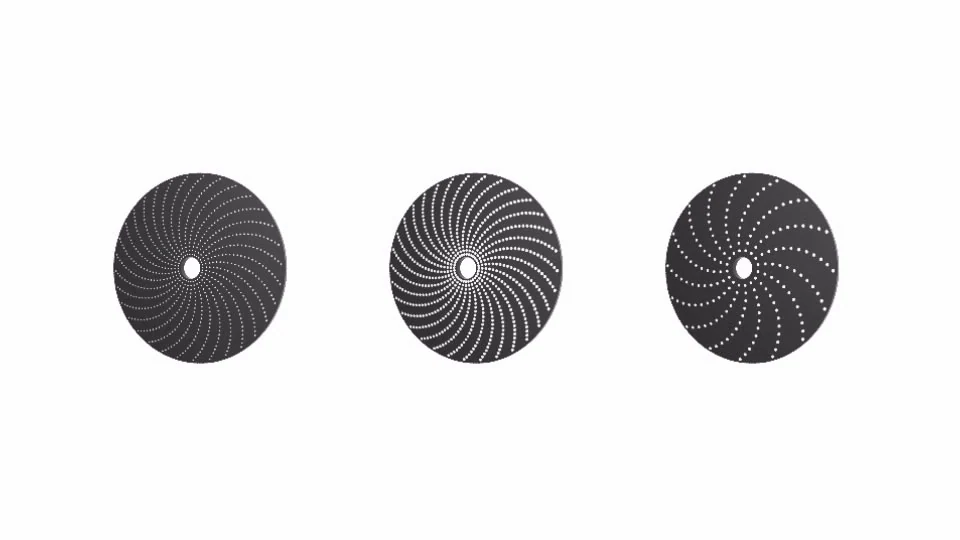
40 µm disk, 60 µm disk, 60 µm DSD
Spinning disks available for the Cytation C10 confocal imaging include 60 µm and 40 µm standard disk and a 60 µm deep-sectioning *(DSD) disk.
The three types span a wide range of magnifications and sample types.
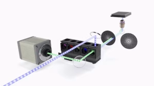 Cytation C10 confocal optical path with two (60 µm and 40 µm) spinning disk, confocal filter cubes and Hamamatsu scientific CMOS camera. Two spinning disk pinhole sizes offer users confocality over a wide range of magnifications. The available 60 µm deep-sectioning disk can provide greater clarity when imaging thick samples.
When optical sectioning is useful
Confocal microscopy is useful for resolving subcellular structures in fluorescently labelled cells and for examining selected depths in spheroids, organoids and tissue sections. Its advantage depends on the amount of background fluorescence and the depth and transparency of the specimen. Thin cell monolayers with little out-of-focus signal may be measured efficiently using widefield fluorescence microscopy.
What determines image quality
Objective numerical aperture, wavelength, pinhole geometry, sampling and refractive-index matching all affect the result. Optical section thickness is different from imaging depth: a thinner section does not mean that light penetrates further. Confocal microscopy also remains diffraction-limited unless combined with a separate super-resolution approach.
Limitations in live and thick samples
Scattering and optical aberrations can limit useful imaging depth, even with confocal detection. In spinning disk systems, light can reach neighbouring pinholes and increase background, an effect known as pinhole cross-talk. Disk design and specimen properties influence its severity. [2] Fluorescence excitation can also cause photobleaching and phototoxicity. Acquisition settings should therefore provide sufficient signal for analysis while limiting illumination and unnecessary Z-sections.
Common questions
Is confocal microscopy always better than widefield imaging?
The appropriate method depends on the sample. Confocal detection is valuable when out-of-focus fluorescence limits contrast; widefield imaging is often efficient for thin specimens and large-area screening.
Does a confocal Z-stack measure the whole organoid?
Only if the acquisition covers the relevant volume and sufficient signal is recovered throughout it. Loss of signal at depth can bias measurements towards accessible regions.
Did you know that the BioTek Cytation C10 combines widefield and confocal microscopy?
BioTek Cytation C10 Combines spinning disk confocal and widefield imaging, with multimode reading available in suitable configurations. Objectives and disk options depend on configuration.
Need help selecting a platform for your assay? Contact BioSPX to discuss the sample, readout and configuration you need.