
 EN
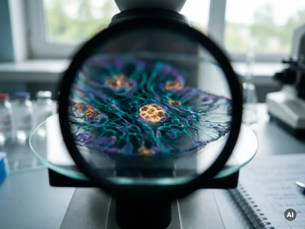
Spatial context is important for understanding molecular mechanisms because the location of molecules, cells, and structures within a biological system directly influences how those molecules behave, interact, and function. Biology is inherently spatial: a gene expressed in one region of a tissue can drive a completely different outcome than the same gene expressed elsewhere. The sections below explore how spatial organization shapes research across cellular biology, disease investigation, and the technologies that make this level of analysis possible.
How does spatial organization shape cellular function?
Spatial organization shapes cellular function by determining which molecules are physically close enough to interact, which signals a cell receives from its neighbors, and how a cell’s position within a tissue influences its identity and behavior. Cells do not operate in isolation: their function is inseparable from their structural context.
Within a single cell, the compartmentalization of organelles ensures that biochemical reactions occur in the right place at the right time. Transcription happens in the nucleus, translation on ribosomes distributed across the cytoplasm, and energy production in the mitochondria. Disrupting this internal spatial order, as occurs in many disease states, can impair cellular function even when the underlying genetic sequence remains unchanged.
At the tissue level, the spatial organization of cells determines which cell types are adjacent to one another, how nutrients and signals are distributed, and how structural gradients are maintained. The architecture of a tissue, whether it is a liver lobule, a layer of the cerebral cortex, or a tumor microenvironment, encodes biological information that is simply not present in a dissociated cell population. Understanding molecular mechanisms therefore requires preserving and mapping this spatial information.
What is lost when spatial context is removed from molecular data?
When spatial context is removed from molecular data, researchers lose information about where within a tissue a particular gene is expressed, which cell types are neighbors, and how local microenvironments influence molecular states. Bulk sequencing and even single-cell approaches that require tissue dissociation average or anonymize this positional information entirely.
Dissociation-based methods treat every cell as an equal unit regardless of its original location. This means that a rare cell population sitting at the edge of a tumor, exposed to hypoxic conditions and adjacent to immune infiltrates, is analyzed in the same pool as a morphologically similar cell in a well-vascularized core. The molecular differences between these two cells may be profound, but without spatial context, the distinction is invisible.
The loss of tissue architecture also removes information about gradients, boundaries, and zonation patterns. Many biological processes, including stem cell niche maintenance, immune cell trafficking, and metabolic zonation in the liver, are defined precisely by spatial gradients of signaling molecules. Stripping away that architecture collapses a three-dimensional biological reality into a flat, context-free data set.
How does spatial context reveal cell-to-cell communication?
Spatial context reveals cell-to-cell communication by showing which cells are physically positioned to exchange signals through direct contact or short-range secreted factors. Without knowing which cells are neighbors, it is impossible to determine whether an observed signaling relationship is biologically meaningful or a computational artifact.
Many signaling pathways operate over very short distances. Juxtacrine signaling, where a ligand on one cell surface binds a receptor on an adjacent cell, requires direct physical proximity. Paracrine signals, while able to travel slightly further, still operate within defined spatial boundaries set by diffusion rates and tissue structure. Spatial transcriptomics and related spatial omics approaches allow researchers to map ligand-receptor co-expression patterns in their native tissue context, making it possible to infer which communication events are actually occurring in situ.
This has particular relevance for understanding how tumor cells recruit supporting stromal cells, how neurons form functional circuits, and how immune cells coordinate responses within inflamed tissue. Identifying these spatial communication patterns can help researchers generate more precise hypotheses about the molecular mechanisms driving these processes.
What are the main spatial biology technologies used today?
The main spatial biology technologies used today include spatial transcriptomics, spatial proteomics, multiplexed imaging approaches, and in situ sequencing methods. Each captures a different layer of molecular information while preserving the positional coordinates of the data within intact tissue sections.
Key spatial biology approaches currently in use include:
- Spatial transcriptomics: Maps gene expression across tissue sections by capturing mRNA at spatially barcoded positions, enabling researchers to link transcriptional profiles to specific tissue regions
- In situ sequencing and hybridization: Detect individual RNA molecules directly within tissue without dissociation, preserving single-cell and subcellular resolution
- Multiplexed immunofluorescence and imaging mass cytometry: Simultaneously visualize dozens of proteins within tissue sections, revealing the spatial distribution of cell types and functional markers
- Spatial proteomics: Extends proteomic profiling to intact tissue, linking protein abundance and modification states to tissue location
- 3D imaging and light-sheet microscopy: Provide volumetric views of intact organs or organoids, enabling spatial analysis across all three dimensions
In 2026, these technologies are increasingly being combined into integrated spatial omics workflows, allowing researchers to co-register transcriptomic, proteomic, and morphological data within the same tissue section. This multimodal approach gives a far richer picture of molecular mechanisms than any single method alone.
How does spatial context advance drug discovery and disease research?
Spatial context advances drug discovery and disease research by revealing how molecular alterations are distributed across diseased tissue, which cell populations drive pathological processes, and how the tissue microenvironment influences cellular behavior. This level of detail supports the identification of more precise research targets and more relevant experimental models.
In cancer research, spatial omics approaches have shown that tumors are far from uniform. Different regions within the same tumor can harbor distinct transcriptional programs, immune infiltration patterns, and metabolic states. Understanding this intratumoral heterogeneity at the spatial level may help researchers investigate why certain cell populations are more resistant to experimental interventions and which microenvironmental factors contribute to that resistance.
In neurological and inflammatory disease research, spatial transcriptomics is being used to map how gene expression changes in specific brain regions or tissue compartments during disease progression. This can reveal early regional changes that are invisible in bulk tissue analysis, potentially supporting the identification of new research questions and mechanistic hypotheses.
For researchers in Belgium, the Netherlands, and Luxembourg working in pharmaceutical R&D, biotech, or academic settings, integrating spatial biology into discovery workflows represents a meaningful methodological advance. The ability to study molecular mechanisms within their native tissue architecture improves the biological relevance of experimental findings.
What imaging and microscopy systems support spatial biology workflows?
Imaging and microscopy systems that support spatial biology workflows include high-content fluorescence microscopes, confocal systems, light-sheet microscopes, and automated slide scanning platforms. These instruments provide the spatial resolution, multiplexing capacity, and throughput needed to generate and analyze spatially resolved molecular data at scale.
The choice of imaging system depends on the spatial resolution required, the number of markers being detected, and whether the sample is a thin tissue section, a whole cleared organ, or a three-dimensional organoid model. For spatial transcriptomics workflows based on fluorescence in situ hybridization, high-sensitivity widefield or confocal systems capable of detecting individual RNA molecules are typically required. For multiplexed protein imaging, systems with broad spectral flexibility and automated stage control support the sequential or simultaneous detection of many markers.
Organoid models are increasingly used as spatially relevant systems for studying molecular mechanisms in three dimensions. Imaging organoids requires instruments capable of capturing volumetric data with sufficient depth penetration, making light-sheet microscopy and spinning disk confocal systems particularly well suited to these workflows. As organoid complexity increases, the demand for imaging systems that can handle live, three-dimensional samples over extended time periods continues to grow.
Automated image analysis and data management are equally important. The data volumes generated by spatial imaging workflows are substantial, and researchers need analysis pipelines capable of registering images, segmenting cells, and linking spatial coordinates to molecular readouts in a reproducible way. Exploring cellular analysis solutions that integrate imaging with downstream data workflows is an important step in building a functional spatial biology setup.
How BioSPX supports spatial biology research
BioSPX supports researchers in Belgium, the Netherlands, and Luxembourg who are building or expanding spatial biology workflows. As an independent life science distributor with deep expertise in imaging, microscopy, and cellular analysis, BioSPX helps laboratories identify the right instruments for their specific spatial research needs.
BioSPX’s support for spatial biology includes:
- Expert guidance on imaging and microscopy systems suited to spatial transcriptomics, multiplexed protein imaging, and organoid analysis
- Advice on instrument configurations and experimental setup from product specialists with application-level knowledge
- Installation, user training, and after-sales support to ensure instruments perform reliably within established workflows
- Access to innovative solutions such as the Orgonex RPMotion bioreactor and the Discover Nestor automation system, which support spatially relevant three-dimensional research models
If you are developing a spatial biology workflow or looking to expand your imaging capabilities, contact BioSPX to speak with a specialist about the right solution for your laboratory.
Frequently Asked Questions
How do I know which spatial biology technology is the right fit for my research question?
The best starting point is to define what molecular layer you need to interrogate — transcriptomic, proteomic, or both — and at what resolution. For example, if subcellular RNA localization is critical, in situ hybridization-based methods offer single-molecule resolution, while array-based spatial transcriptomics is better suited for broad tissue-wide gene expression mapping. Consulting with an application specialist before committing to a platform can save significant time and resources, especially when your workflow involves complex sample types like organoids or cleared tissue.
Can spatial biology approaches be integrated with existing single-cell RNA sequencing (scRNA-seq) data?
Yes, and this integration is increasingly common in spatial omics workflows. Spatial transcriptomics data can be computationally deconvolved using reference scRNA-seq datasets to assign cell type identities to spatial spots or regions, combining the resolution of single-cell profiling with the positional information of spatial data. Tools such as RCTD, Tangram, and Cell2location are widely used for this purpose. The key requirement is that both datasets are derived from comparable tissue or biological conditions to ensure meaningful integration.
What are the most common technical challenges when setting up a spatial transcriptomics workflow, and how can they be avoided?
Tissue quality is the single most critical variable — RNA degradation from suboptimal fixation, freezing, or sectioning can severely compromise data quality before any instrument is even involved. Ensuring consistent cryosectioning thickness, using RNase-free reagents throughout, and optimizing permeabilization conditions for your specific tissue type are essential early steps. Working with experienced application support during the initial setup phase, rather than troubleshooting independently, is one of the most effective ways to avoid costly experimental failures.
Is spatial biology only relevant for cancer research, or does it apply broadly across disease areas?
Spatial biology is broadly applicable across virtually every disease area where tissue architecture and cellular microenvironment play a role — which is most of them. Beyond oncology, active spatial research is being conducted in neurodegeneration, autoimmune and inflammatory diseases, cardiovascular biology, developmental disorders, and infectious disease. Any research question that involves understanding how molecular states vary across tissue regions, how cell types communicate in situ, or how disease alters tissue organization can benefit from a spatial approach.
How much computational infrastructure is needed to analyze spatial omics data?
Spatial omics datasets are substantially larger and more complex than standard sequencing or imaging outputs, so computational infrastructure is a genuine consideration. At minimum, researchers need access to high-memory computing environments and familiarity with analysis frameworks such as Seurat, Squidpy, or MERFISH-specific pipelines for transcriptomic data, and platforms like HALO or QuPath for image-based spatial analysis. Cloud computing resources and institutional HPC clusters are increasingly used to handle the data volumes involved, and planning your data management and analysis strategy before generating data is strongly recommended.
Can organoids reliably replicate the spatial complexity of native tissue for spatial biology studies?
Organoids recapitulate many aspects of native tissue spatial organization, including self-organized cellular compartments, gradients, and cell-type diversity, making them valuable and tractable models for spatial biology. However, they do not fully reproduce the vascular networks, immune infiltration, or mechanical cues present in vivo, which are factors that can meaningfully influence molecular states. The most robust research strategies use organoids alongside primary tissue validation, leveraging the experimental accessibility of organoids while confirming key spatial findings in native tissue contexts.
What should I prioritize when selecting an imaging system for a multiplexed spatial proteomics workflow?
Spectral flexibility and sensitivity are the primary imaging considerations for multiplexed protein detection, as you need to resolve a large number of markers without significant spectral bleed-through. Automated stage control and reproducible slide registration are equally important for workflows involving sequential staining cycles, where precise image alignment across rounds is essential for accurate co-localization analysis. Throughput and compatibility with your downstream image analysis software should also factor into the decision, particularly if you are planning to process large tissue cohorts at scale.
Related Articles
- How does in situ analysis preserve cellular and tissue information?
- How does angiogenesis support tumor growth at the cellular level?
- How do researchers distinguish cytostatic from cytotoxic drug effects in vitro?
- What is the difference between 2D and 3D tumor cell culture models?
- How does hypoxia affect cancer cell survival and proliferation?
This content was generated with the help of AI and it may contain mistakes