
 EN
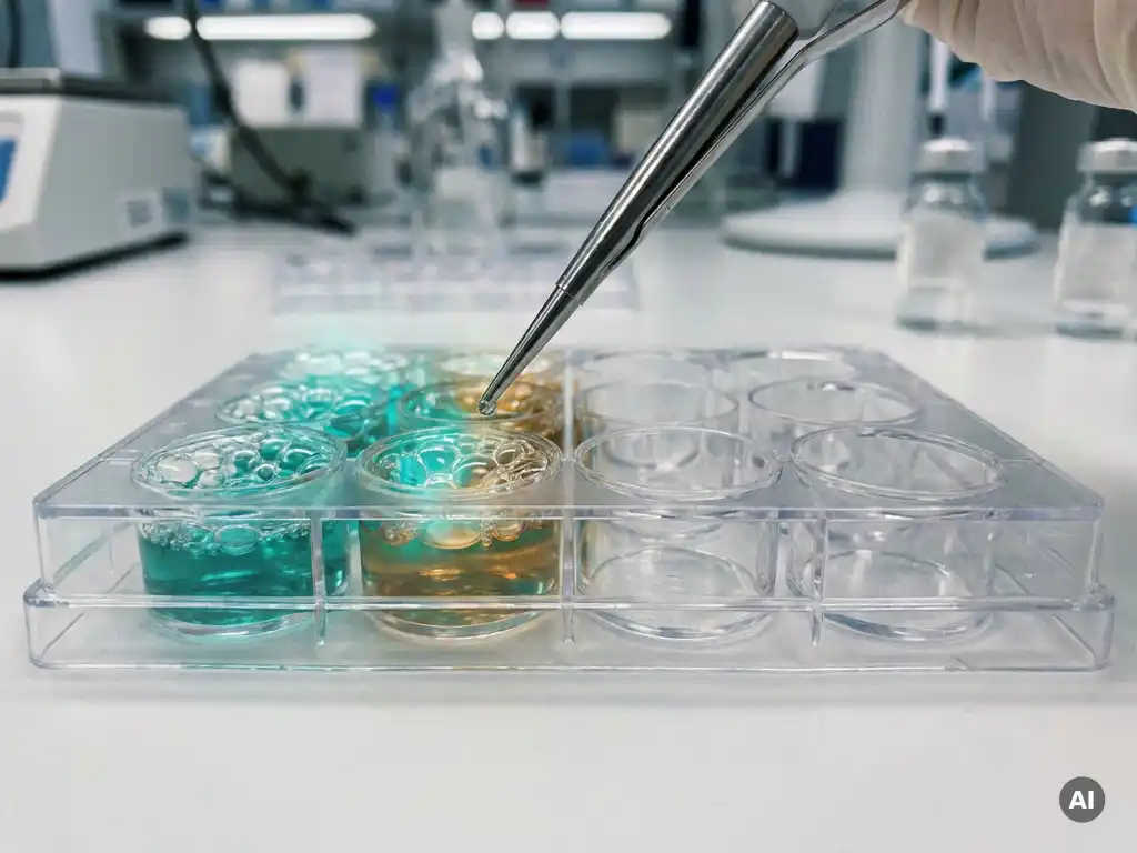
Measuring protein function is more informative than measuring protein presence because presence alone tells you a protein exists, while function tells you whether it is actually doing anything biologically meaningful. A protein can be abundant in a sample yet completely inactive due to post-translational modifications, misfolding, or inhibitory interactions. For researchers in life sciences, this distinction shapes experimental conclusions and the reliability of downstream data. The sections below address the most common questions about functional protein measurement and when it matters most in laboratory practice.
What does measuring protein function actually reveal?
Measuring protein function reveals the biological activity of a protein within a given cellular or biochemical context. Rather than confirming that a protein is present at a certain concentration, functional measurement determines whether that protein is performing its intended role, such as catalyzing a reaction, binding a target molecule, activating a signaling cascade, or regulating gene expression.
This distinction matters because proteins operate within complex networks. A kinase, for example, may be expressed at high levels but remain inactive if its upstream activator is absent. An enzyme may be present in abundance but unable to reach its substrate due to compartmentalization. Functional assays capture these nuances by measuring outputs such as enzyme velocity, receptor binding affinity, signal transduction efficiency, or downstream effector activation.
For researchers working in cellular and molecular biology, functional data provides a more complete picture of biological state. It can reveal whether a pathway is truly active, whether a perturbation has had the intended effect, and whether observed phenotypes are mechanistically connected to a specific protein’s activity.
Why can high protein levels still mean low biological activity?
High protein levels can coexist with low biological activity because protein abundance and protein function are regulated by different mechanisms. Expression levels reflect how much protein has been synthesized, but activity depends on factors including post-translational modifications, protein-protein interactions, subcellular localization, and the availability of cofactors or substrates.
Several common scenarios illustrate this disconnect:
- Post-translational modification: Phosphorylation, ubiquitination, acetylation, and glycosylation can activate or silence a protein regardless of its concentration.
- Inhibitory binding partners: Endogenous inhibitors or competing proteins can block active sites even when total protein levels are high.
- Misfolding or aggregation: A protein that has not folded correctly cannot perform its function, even if it is detectable by antibody-based assays.
- Compartmentalization: A protein present in the wrong cellular compartment may be isolated from its substrates or binding partners.
- Zymogen or precursor forms: Many enzymes are synthesized as inactive precursors that require proteolytic cleavage to become active.
This is why protein biomarker studies that rely solely on quantification methods can sometimes yield misleading conclusions. The biologically relevant question is not how much protein is present, but how much is functionally engaged.
How do functional protein assays work in practice?
Functional protein assays work by measuring a biological output that depends on the protein’s activity rather than its physical presence. The specific readout varies by protein type, but common approaches include enzymatic activity assays, binding assays, reporter gene assays, and cell-based assays that capture downstream signaling events.
In an enzymatic activity assay, the rate at which a substrate is converted to a product serves as a direct measure of enzyme function. Fluorescent or luminescent substrates are frequently used because they allow continuous, real-time monitoring without the need to stop the reaction at fixed time points.
Cell-based assays take a broader approach by measuring how a protein’s activity affects cellular behavior. This can include changes in proliferation, differentiation, secretion, or reporter gene expression. These assays are particularly valuable when the goal is to understand protein function within a physiologically relevant context rather than in an isolated biochemical system.
Binding assays, such as surface plasmon resonance or bead-based interaction studies, measure the affinity and kinetics with which a protein interacts with its partners. This type of functional measurement is especially relevant in drug discovery workflows, where understanding how a candidate compound affects protein-protein or protein-ligand interactions is a key research objective.
What’s the difference between protein quantification and protein activity assays?
Protein quantification measures how much of a protein is present in a sample, while protein activity assays measure what that protein is doing. The two approaches answer fundamentally different questions and are optimized for different experimental purposes.
Protein quantification methods, including Western blotting, ELISA, and mass spectrometry-based proteomics, use antibodies or physicochemical properties to detect and count protein molecules. They are well-suited for comparing expression levels across conditions, validating knockdown or overexpression, or identifying proteins in complex mixtures.
Protein activity assays, by contrast, measure a functional output. They require the protein to be in a biologically active state and often depend on intact enzymatic machinery, appropriate cofactors, and suitable assay conditions. The readout reflects what the protein is capable of doing under the tested conditions, not simply how many molecules are present.
In practice, the most robust experimental designs combine both approaches. Quantification confirms that changes in activity are not simply the result of differences in expression, while activity assays confirm that expression differences translate into meaningful functional changes. Together, they support stronger, more reproducible conclusions in functional proteomics research.
When should researchers choose functional over presence-based measurements?
Researchers should prioritize functional measurements over presence-based detection when their scientific question centers on biological activity, pathway engagement, or the mechanistic effect of an experimental perturbation. Presence-based methods are sufficient when the goal is simply to confirm expression, but they fall short when activity is the key variable.
Functional assays are particularly appropriate in the following research contexts:
- Enzyme characterization: When studying how a modification, mutation, or compound affects catalytic efficiency.
- Signaling pathway analysis: When determining whether a pathway is active, not merely whether its components are expressed.
- Drug target engagement studies: When confirming that a compound inhibits or activates a specific protein’s function in a cellular system.
- Biomarker validation: When a protein biomarker is being evaluated for its functional relevance rather than its abundance alone.
- Quality control of recombinant proteins: When verifying that a produced protein is correctly folded and active before use in downstream experiments.
In settings where both expression and activity data are needed, functional assays should be designed in parallel with quantification methods rather than treated as an alternative. This is especially relevant in pharmaceutical and biotech research environments, where decision-making depends on mechanistic evidence rather than correlative expression data.
What instruments are used to measure protein function in the lab?
The instruments used to measure protein function depend on the assay format, the type of readout, and the throughput requirements of the experiment. Several instrument categories are central to functional protein measurement in modern life science laboratories.
Multimode readers are among the most versatile instruments for protein function measurement. They can detect fluorescence intensity, luminescence, absorbance, and time-resolved fluorescence in microplate format. This makes them suitable for enzymatic activity assays, FRET-based binding assays, reporter gene assays, and cell viability measurements that serve as functional endpoints. For laboratories running multiple assay types, a multimode reader offers the flexibility to consolidate several measurement needs into a single platform.
Imaging systems, including high-content imaging platforms and fluorescence microscopes, are used when spatial information about protein activity is needed. These instruments can capture subcellular localization of active signaling molecules, translocation events, or changes in protein distribution that accompany functional shifts.
Liquid handling systems support high-throughput functional assay workflows by automating reagent addition, serial dilutions, and plate preparation. Consistent liquid handling is critical for reproducible enzymatic and cell-based assays, where small volume errors can significantly affect measured activity values.
Microplate washers and dispensers play a supporting role in assay formats that require washing steps, such as bead-based binding assays or cell-based assays with multiple reagent additions. Precise dispense volumes and wash efficiency directly affect the signal-to-noise ratio and reproducibility of functional readouts.
How BioSPX supports protein function measurement in the Benelux
BioSPX provides life science laboratories across Belgium, the Netherlands, and Luxembourg with the instruments and expertise needed to move from protein detection toward meaningful functional analysis. Whether a laboratory is establishing a new functional assay workflow or scaling existing measurements to higher throughput, BioSPX offers:
- A broad portfolio of multimode readers, imaging systems, liquid handling solutions, and microplate dispensers suited to enzymatic, cell-based, and binding assays
- Product specialists who advise on instrument selection, assay compatibility, and experimental configuration
- Installation, user training, and responsive maintenance support to ensure instruments perform reliably over time
- ISO 9001-certified service standards with offices in the Netherlands and Belgium
If you are evaluating instruments for functional protein assays or want guidance on which platform best fits your research workflow, contact BioSPX to speak with a specialist.
Frequently Asked Questions
Can I use the same sample for both protein quantification and functional activity assays?
In many cases, yes, but sample preparation conditions need to accommodate both measurement types. Quantification methods such as Western blotting or ELISA are generally tolerant of denaturing conditions, while functional activity assays require the protein to remain in its native, active conformation. If you plan to run both assay types from a single sample, prepare your lysate or extract under non-denaturing conditions and confirm that your buffer composition is compatible with the activity assay readout. Dividing the sample into aliquots immediately after extraction helps preserve activity and avoids repeated freeze-thaw cycles that can degrade enzymatic function.
What are the most common mistakes researchers make when setting up a functional protein assay for the first time?
The most frequent pitfalls include using assay conditions that are incompatible with the protein’s active state, such as incorrect pH, temperature, or missing cofactors, and failing to validate the linear range of the assay before interpreting results. Another common error is not including appropriate controls, specifically a positive control with known activity and a negative control where the protein is inactivated or absent. Without these, it is difficult to distinguish true biological signal from background noise or assay artifacts. Running a substrate titration and a protein concentration curve before the main experiment will save significant time and reagents.
How do I know if a decrease in measured activity reflects a real biological change or a technical issue with my assay?
The first step is to check your positive control: if a sample with known, stable activity shows a similar drop, the issue is likely technical rather than biological. Common technical causes include reagent degradation, pipetting inconsistencies, substrate depletion in high-activity samples, or instrument calibration drift. If your controls perform as expected, replicate the result across independent biological replicates and, where possible, confirm the activity change using an orthogonal assay format. Pairing activity data with quantification data, such as a Western blot, also helps determine whether the change in activity correlates with a change in protein levels or reflects a true shift in specific activity.
Are cell-based functional assays always more physiologically relevant than cell-free enzymatic assays?
Cell-based assays offer greater physiological context because they preserve the cellular environment, including endogenous binding partners, compartmentalization, and regulatory networks, but this complexity also introduces more variables that can be difficult to control or interpret. Cell-free enzymatic assays, by contrast, offer precision and mechanistic clarity, making them ideal for characterizing kinetic parameters, testing compound potency, or isolating the effect of a specific modification on catalytic activity. The right choice depends on your scientific question: if you need to understand intrinsic protein function or screen compounds efficiently, a cell-free assay is often preferable; if you need to confirm that activity translates into a cellular response, a cell-based format is more appropriate.
How do post-translational modifications affect the outcome of functional assays, and should I account for them in my assay design?
Post-translational modifications (PTMs) can dramatically alter a protein’s activity, substrate specificity, or interaction profile, and if not accounted for, they can lead to misleading assay results. For example, if you are working with a recombinant protein expressed in a bacterial system, it may lack the phosphorylation or glycosylation events present in the native human protein, resulting in different activity levels than what would be observed in a physiological setting. When designing functional assays, consider whether your protein source reflects the modification state relevant to your biological question, and where possible, use phospho-specific antibodies or mass spectrometry to characterize the modification status of your protein alongside the functional measurement.
What throughput options are available for functional protein assays, and how do I scale up without losing data quality?
Functional protein assays can be scaled from low-throughput single-well formats to high-throughput 384- or 1536-well microplate formats, depending on the assay type and available instrumentation. The key to maintaining data quality during scale-up is consistent liquid handling: small volume errors in high-density plate formats have a proportionally larger impact on measured activity than in standard 96-well plates. Automated liquid handling systems and microplate dispensers are strongly recommended for high-throughput workflows. Additionally, validating assay performance metrics such as the Z’-factor, signal-to-background ratio, and coefficient of variation at the intended scale before committing to a full screen will help ensure reproducibility and data integrity.
How should I approach selecting a multimode reader for functional protein assays if my laboratory runs several different assay types?
Start by mapping the detection modalities required across all your current and planned assay types, such as fluorescence intensity, luminescence, absorbance, time-resolved fluorescence, or fluorescence polarization, and prioritize an instrument that covers the broadest range without compromising sensitivity on any single mode. Consider plate format compatibility, injection capabilities for kinetic assays, and whether the software supports the data analysis workflows your team already uses. Consulting with an instrument specialist who understands your specific assay portfolio, rather than selecting based on specifications alone, can prevent costly mismatches between instrument capability and experimental need.
Related Articles
- What is clonal evolution and how does it shape tumor heterogeneity?
- How can flow cytometry complement live-cell imaging in treatment response studies?
- How do cancer cells develop resistance to treatment over time?
- What is targeted therapy and how is it different from chemotherapy?
- What is the difference between curative and palliative cancer treatment?
This content was generated with the help of AI and it may contain mistakes