
 EN
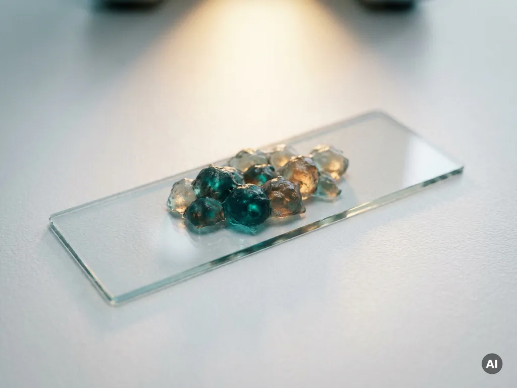
Cancer cells behave differently across repeated experiments primarily because of their inherent biological variability, compounded by changes that accumulate during routine laboratory handling. Cell populations are not uniform; they contain subpopulations with distinct genetic and epigenetic profiles that shift over time and in response to culture conditions. Understanding the specific drivers of this variability is essential for any researcher aiming to generate reproducible, meaningful data.
The sections below address the most common questions researchers ask when they notice inconsistency in cancer cell experiments, from the biology of heterogeneous populations to practical strategies for tightening experimental control.
Why do cancer cells change behavior between experiments?
Cancer cells change behavior between experiments because they are biologically unstable populations subject to continuous genetic drift, selective pressure, and environmental influence. Unlike normal differentiated cells, cancer cell lines often carry chromosomal instability that drives ongoing mutation and phenotypic divergence, meaning the cells used in one experiment may differ meaningfully from those used weeks or months later.
This instability is not random noise; it reflects the underlying biology of cancer itself. Tumor cells evolve under selective pressure, and in culture that pressure comes from the laboratory environment: the composition of the growth medium, oxygen tension, temperature fluctuations, and competitive dynamics within the flask. Cells that proliferate fastest under those specific conditions gradually dominate the population, shifting the overall behavior of the line.
For researchers in the Benelux working across oncology, pharmaceutical R&D, or biotech, this means that cancer cell variability is not simply a technical inconvenience. It is a biological phenomenon that must be actively managed through consistent culture practices and rigorous experimental design.
What is phenotypic heterogeneity in cancer cell populations?
Phenotypic heterogeneity in cancer cell populations refers to the coexistence of cells with distinct functional characteristics within the same culture, even when those cells share a common origin. These differences in morphology, proliferation rate, drug sensitivity, and gene expression arise from genetic mutations, epigenetic variation, and stochastic differences in gene regulation.
In practical terms, this means that a flask of cancer cells is not a single uniform entity. It is a dynamic ecosystem of subpopulations. Some subpopulations may be more proliferative, others more quiescent, and others may express surface markers or signaling proteins at levels that differ substantially from the majority.
This heterogeneity has direct consequences for experimental reproducibility in cancer research. When researchers seed cells for an assay, the proportion of each subpopulation in that seeding aliquot may vary depending on how the cells were passaged, how confluent the flask was, and how long the culture has been maintained. These proportional shifts can produce measurable differences in assay outcomes even when all other protocol steps remain identical.
Recognizing phenotypic heterogeneity as a structural feature of cancer cell populations, rather than a sign of experimental error, is the first step toward designing experiments that account for it appropriately.
How does cell passaging affect experimental results?
Cell passaging affects experimental results by progressively altering the genetic composition, growth characteristics, and stress responses of a cancer cell line. Each passaging event introduces selective pressure: cells that detach easily, recover quickly, and adapt to the new seeding density are more likely to dominate subsequent passages, gradually changing the population’s overall behavior.
The effect is cumulative. Early-passage cells and high-passage cells of the same nominal line can exhibit substantially different proliferation rates, morphology, receptor expression, and sensitivity to experimental stimuli. This is a well-recognized source of cell culture variability that affects reproducibility across time points and between laboratories.
Key passaging-related factors that contribute to variability include:
- Passage number at the time of the experiment
- Inconsistent split ratios between passages
- Variable recovery time after thawing from cryopreservation
- Differences in the enzyme or dissociation method used to detach cells
- Variation in the duration of enzymatic treatment
Maintaining detailed passage logs and working within a defined passage window are among the most effective ways to reduce this source of experimental drift.
What environmental factors make cancer cells behave inconsistently?
Environmental factors that make cancer cells behave inconsistently include fluctuations in temperature, CO2 concentration, humidity, pH, and media composition. Even small deviations from optimal conditions can alter cell metabolism, signaling pathway activity, and proliferation rate, producing results that are difficult to compare across experiments.
Media-related variables deserve particular attention. The lot number of serum used in growth media can introduce significant biological variability, since serum composition is not standardized between batches. A change in serum lot mid-study can shift baseline proliferation rates or alter the expression of surface markers, making it appear as though the cells themselves have changed when the variable is actually the medium.
Incubator performance is another underappreciated source of inconsistency. Shared incubators that are frequently opened introduce temperature and CO2 fluctuations, and contamination risk increases with the number of users. Spatial positioning within the incubator can also matter, as temperature gradients across shelves may affect evaporation rates and edge effects in multiwell plates.
Beyond the incubator, the timing and handling of cells during an experiment introduce variability. The duration cells spend outside the incubator during media changes or imaging, the temperature of reagents at the point of addition, and the consistency of pipetting technique all contribute to the overall variability profile of a cancer cell experiment.
How can researchers reduce variability in cancer cell experiments?
Researchers can reduce variability in cancer cell experiments by standardizing every controllable element of the culture and assay workflow, using authenticated cell lines, banking early-passage stocks, and applying consistent environmental conditions across all experimental runs.
A structured approach to reducing cancer research reproducibility challenges typically involves the following steps:
- Authenticate cell lines using short tandem repeat profiling before beginning any study, and retest periodically during long-running projects.
- Establish a master cell bank from low-passage stocks and thaw fresh vials at regular intervals to avoid passage-related drift.
- Standardize media preparation by reserving sufficient serum from a single lot for the duration of a project and testing new lots before switching.
- Document and control passage number by working within a defined passage window and discarding cultures that exceed it.
- Minimize manual handling variation by using calibrated pipettes, consistent seeding densities, and standardized timing for all steps.
- Monitor incubator conditions continuously and log any deviations that coincide with experimental runs.
Laboratory automation can play a meaningful role in reducing the human-introduced variability that accumulates across repeated manual handling steps. Automated liquid handling and cell culture systems can apply the same pipetting volumes, timing, and media exchange protocols with a consistency that is difficult to achieve manually, particularly across multi-day experiments.
When should inconsistent cancer cell results prompt a protocol review?
Inconsistent cancer cell results should prompt a protocol review when the same experiment produces statistically divergent outcomes across biological replicates that share the same passage number, media lot, and culture conditions. Occasional variation is expected, but systematic inconsistency that follows a pattern, for example, results drifting over time or differing between operators, signals a controllable source of variability that warrants investigation.
Specific triggers that justify a formal protocol review include:
- Results that are reproducible within a single operator’s hands but not between operators
- Outcomes that shift after a reagent lot change, equipment service, or incubator relocation
- Morphological changes or unexpected growth kinetics in a cell line that has been in use for an extended period
- Assay signals that fall outside the expected range without a clear experimental explanation
A structured root cause analysis should examine passage history, reagent lot records, equipment calibration logs, and environmental monitoring data before concluding that the biology itself is responsible for the inconsistency. In many cases, the source of cancer cell behavior variability can be traced to a procedural or environmental factor that is fully addressable.
Researchers who build regular protocol audits into their experimental calendar, rather than waiting for a reproducibility failure to trigger a review, are better positioned to catch drift early and maintain data quality over the course of a project.
How BioSPX supports reproducibility in cancer cell research
For research laboratories in the Benelux looking to address the sources of cancer cell variability described above, BioSPX offers instrumentation and expertise specifically relevant to cell culture consistency and experimental standardization. The portfolio includes solutions that directly target the controllable variables that drive inconsistent results:
- Automated cell culture systems that reduce manual handling variability across feeding, media exchange, and passage workflows
- Liquid handling solutions that apply consistent volumes and timing across multiwell plate formats
- Imaging and microscopy systems for monitoring cell morphology and growth kinetics in a standardized, quantifiable way
- Multimode readers for reproducible, high-quality assay readouts across experimental runs
- Expert guidance from product specialists who advise on experimental setup, system configuration, and workflow optimization
BioSPX also provides installation, user training, and responsive maintenance support to ensure that instruments perform consistently over time. If you are investigating ways to improve reproducibility in your cancer cell research, get in touch with our team to discuss which solutions are best suited to your laboratory’s specific workflow and research goals.
Frequently Asked Questions
How do I know if my cancer cell line has drifted too far to be usable?
Key warning signs include morphological changes visible under the microscope, a significant shift in doubling time compared to your baseline records, unexpected resistance or sensitivity to a treatment that previously produced consistent results, and STR profiling results that no longer match the reference profile. If any of these indicators appear, it is advisable to discard the current culture and thaw a fresh vial from your master cell bank rather than continuing with a compromised population.
What is the recommended passage window for maintaining consistent cancer cell behavior?
While the optimal passage window varies by cell line, most laboratories working with cancer cell lines aim to keep experiments within a range of 10–20 passages from the original thaw, and many guidelines recommend not exceeding passage 30 from the authenticated stock. The most important practice is to define and document your own passage window based on your specific cell line’s stability, test behavior at the upper limit of that window, and always record the passage number in your experimental metadata so that it can be used as a variable during data analysis.
Can mycoplasma contamination explain inconsistent cancer cell results even without visible signs of infection?
Yes, and this is one of the most commonly overlooked sources of experimental inconsistency. Mycoplasma contamination does not cause visible turbidity or obvious cell death, but it can significantly alter cell proliferation rates, cytokine secretion, gene expression profiles, and drug sensitivity — all of which directly affect assay outcomes. Routine mycoplasma testing, ideally every one to two months and after any new cell line is introduced to the laboratory, should be a standard part of any reproducibility management strategy.
Is it better to use single-cell clones to reduce heterogeneity, and what are the trade-offs?
Single-cell cloning can reduce phenotypic heterogeneity within a working population and is a valid strategy for specific applications such as CRISPR editing validation or stable overexpression studies. However, clonal populations are not representative of the parental line’s heterogeneity, and clonal expansion itself introduces selection bias, meaning the resulting clone reflects only one subset of the original population’s biology. For experiments intended to model tumor biology broadly, working with a well-maintained, authenticated polyclonal population is generally more appropriate than relying on clonal derivatives.
How should I handle the transition between serum lots to minimize disruption to ongoing experiments?
The best practice is to reserve enough serum from a single validated lot to cover the full duration of a project before it begins. When a lot transition is unavoidable, run a parallel comparison by culturing your cells simultaneously in the old and new lots for at least two to three passages, measuring key functional parameters such as proliferation rate, viability, and any assay-specific readouts. Only switch to the new lot once you have confirmed that the functional performance of your cells is equivalent, and document the lot transition date in your experimental records.
What is the minimum documentation a lab should keep to troubleshoot cancer cell variability effectively?
At a minimum, every experiment should be linked to records of the cell line identity and STR authentication date, passage number, thaw date and source vial, media and serum lot numbers, incubator ID and any logged deviations, and the operator who performed key steps. This metadata is what makes it possible to identify the root cause of a reproducibility failure after the fact — without it, troubleshooting becomes guesswork. Embedding this documentation into a standardized lab notebook template or electronic lab notebook system is the most reliable way to ensure it is captured consistently.
At what point does it make practical sense to consider laboratory automation for cancer cell culture?
Automation becomes particularly valuable when experiments involve high-throughput screening across many conditions, when multi-day culture protocols require precise and consistent media exchange timing, or when reproducibility failures have been traced back to manual handling variability between operators. Even in smaller labs, targeted automation of high-impact steps — such as liquid dispensing for plate-based assays — can meaningfully reduce variability without requiring a full automated cell culture system. A practical starting point is to identify the two or three manual steps in your workflow that are most difficult to perform consistently, and evaluate whether an automated solution addresses those specific bottlenecks.
Related Articles
- How does FRET measure molecular proximity below the resolution limit of conventional microscopy?
- How does photobleaching affect fluorescence-based molecular interaction measurements?
- What is the tumor microenvironment and why does it matter in research?
- How do cancer cells develop resistance to treatment over time?
- What is liquid biopsy and how is it used in oncology?
This content was generated with the help of AI and it may contain mistakes