
 EN
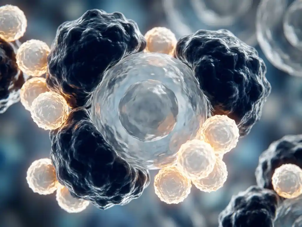
Cancer affects the immune system in two distinct ways: it can directly suppress immune function, and the immune system actively responds to cancer cells, often in a losing battle against the tumor’s evasion strategies. The relationship between cancer and immunity is bidirectional and highly complex. Understanding this interaction is central to modern oncology research and the development of immunological approaches to cancer biology.
How does the immune system normally detect cancer cells?
The immune system detects cancer cells by recognizing abnormal proteins, called neoantigens, displayed on their surface. When a healthy cell mutates and begins behaving abnormally, immune cells, particularly cytotoxic T lymphocytes and natural killer (NK) cells, identify these molecular warning signals and mount a targeted response to eliminate the threat before it can proliferate.
This process is part of a broader framework known as cancer immunosurveillance. The immune system continuously patrols the body, and dendritic cells play a key role by capturing antigens from abnormal cells and presenting them to T cells, effectively activating the adaptive immune response.
Under normal conditions, this surveillance system is remarkably effective. Most nascent tumor cells are eliminated before they can form a detectable mass. The challenge arises when cancer cells accumulate mutations that allow them to evade these detection mechanisms, shifting the balance from immune control to tumor progression.
How do tumors escape immune detection?
Tumors escape immune detection through a process called tumor immune evasion, in which cancer cells acquire characteristics that allow them to hide from or actively disable immune responses. This is one of the most intensively studied areas in cancer immunology research, and it involves multiple overlapping mechanisms that collectively allow malignant cells to persist and grow.
Key evasion strategies include:
- Downregulation of MHC-I molecules: Cancer cells reduce the surface expression of major histocompatibility complex class I proteins, making them harder for cytotoxic T cells to recognize.
- Expression of immune checkpoint ligands: Tumors can express proteins such as PD-L1, which bind to inhibitory receptors on T cells and effectively switch off the immune response.
- Secretion of immunosuppressive cytokines: Molecules such as TGF-beta and IL-10 are released by tumor cells to dampen surrounding immune activity.
- Recruitment of regulatory T cells: Tumors attract immunosuppressive cell types that further inhibit effector immune responses within the tumor site.
- Antigen loss: Over time, cancer cells may lose or mutate the very antigens that initially triggered immune recognition, rendering earlier immune responses ineffective.
These mechanisms do not occur in isolation. Most tumors employ several strategies simultaneously, making immune evasion a multilayered problem that researchers continue to investigate at the cellular and molecular level.
What is the tumor microenvironment and why does it matter?
The tumor microenvironment (TME) is the complex ecosystem surrounding a tumor, composed of cancer cells, immune cells, stromal cells, blood vessels, signaling molecules, and extracellular matrix components. It matters because it actively shapes whether the immune system can mount an effective response, and it is increasingly recognized as a critical determinant of tumor behavior in research models.
Within the TME, immune cells are often present but functionally impaired. T cells may infiltrate the tumor yet fail to kill cancer cells due to local immunosuppressive signals. Tumor-associated macrophages frequently adopt a pro-tumor phenotype, supporting cancer growth rather than opposing it. This hostile environment essentially reprograms immune cells to tolerate, and even support, the tumor.
From a research perspective, modeling and studying the TME has become a major focus. Advanced in vitro systems, including three-dimensional organoid cultures, are being used to replicate the spatial and biochemical complexity of the TME more accurately than traditional two-dimensional cell culture methods. These models allow researchers to study immune-tumor interactions under conditions that more closely reflect what occurs in living tissue.
Does cancer cause a weakened immune system throughout the body?
Yes, cancer can cause systemic immune suppression, meaning the effects on immunity extend beyond the tumor site to affect the broader immune system. The degree of systemic immunosuppression varies considerably depending on cancer type, stage, and the individual’s baseline immune status, but it is a well-documented phenomenon in cancer biology research.
Several mechanisms contribute to this systemic effect. Tumors release soluble factors into circulation that alter the behavior of immune cells throughout the body. In some hematological cancers, the bone marrow, where immune cells are produced, is directly compromised, reducing the overall output of functional lymphocytes and other immune effectors.
Cancer-related systemic immunosuppression also has indirect causes. Nutritional deficits associated with advanced disease, tumor-driven metabolic reprogramming, and the chronic inflammatory state that many cancers induce all contribute to a gradual erosion of immune competence. This is a significant area of interest for researchers studying cancer immunosuppression and its downstream effects on disease progression.
How do cancer treatments affect immune function?
Cancer treatments have varied and sometimes opposing effects on immune function. Some treatments suppress immune activity as a side effect, while others are specifically designed to enhance it. Understanding these effects is a central concern in translational cancer research, where preserving or restoring immune competence is increasingly viewed as integral to research strategy.
Treatments that can suppress immune function
Conventional cytotoxic chemotherapy targets rapidly dividing cells, which includes not only cancer cells but also the proliferating immune cells of the bone marrow and lymphoid tissues. This can result in significant reductions in white blood cell counts, leaving the body temporarily vulnerable. Radiation therapy directed at lymphoid organs or large tissue volumes can have similar effects on local and systemic immune populations.
Treatments that engage or restore immune function
Immunotherapy-based research approaches work in the opposite direction. Checkpoint inhibitor research, CAR-T cell development, and cancer vaccine platforms all aim to reinstate or amplify the immune response to cancer. These approaches do not administer drugs to patients in the clinical sense considered here, but are studied extensively in laboratory and early-phase research settings to understand how the immune system can be redirected against tumor cells.
The interaction between conventional cancer treatment and immune function is an active area of investigation, with researchers exploring combination strategies that minimize immune harm while maximizing anti-tumor activity.
Can the immune system be strengthened to fight cancer?
Research strongly suggests that the immune system can be enhanced to improve its ability to recognize and respond to cancer cells. This is a promising area of investigation, with multiple scientific strategies being explored to boost immune competence, restore exhausted immune cells, and overcome the suppressive signals generated by tumors.
Current research directions include:
- Adoptive cell therapy research: Scientists are investigating how to expand and engineer a patient’s own immune cells outside the body before reintroducing them, with the goal of producing a more potent anti-tumor response.
- Neoantigen-based vaccine research: Personalized approaches that target the unique mutational profile of individual tumors are being studied as a way to prime the immune system more precisely.
- Combination immunological strategies: Research is exploring how pairing different immune-engaging approaches may overcome the redundancy of tumor evasion mechanisms.
- Microbiome research: Emerging evidence suggests that the gut microbiome influences systemic immune tone, and its relationship to cancer immunity is an area of growing scientific interest.
- Organoid and 3D model platforms: Researchers use advanced culture systems to test immune-stimulating strategies in environments that more faithfully replicate tumor biology, enabling more predictive preclinical data.
While no approach can be described as universally effective, the science of immune enhancement in the context of cancer research represents one of the most dynamic frontiers in life sciences today. Researchers in the Benelux region and globally are actively contributing to this evolving body of knowledge.
How BioSPX supports cancer and immune system research
For research teams in the Benelux region studying the relationship between cancer and the immune system, having the right instrumentation and scientific support is essential. BioSPX offers a portfolio of life science instruments specifically suited to the demands of cancer immunology and tumor biology research.
- Organoid and 3D culture systems for modeling the tumor microenvironment with greater biological relevance
- Automated cell culture solutions that reduce manual variability in long-term immune cell and tumor cell culture workflows
- Multimode readers and imaging systems for quantifying immune cell activity, cytokine release, and cellular interactions
- Liquid handling platforms for high-throughput assay preparation in immunological research workflows
- Expert product specialists who advise on instrument selection, experimental setup, and system configuration for specific research applications
BioSPX serves academic laboratories, biotech companies, pharmaceutical R&D teams, and clinical research institutions across Belgium, the Netherlands, and Luxembourg. Whether you are setting up a new immunology research workflow or scaling an existing platform, the team is ready to help you find the right solution. Contact BioSPX to discuss your research needs.
Frequently Asked Questions
What is the difference between 'hot' and 'cold' tumors in immunology research?
In cancer immunology, ‘hot’ tumors are highly infiltrated with active immune cells, particularly T cells, and tend to show more responsiveness to immune-based research interventions. ‘Cold’ tumors, by contrast, have little to no immune cell infiltration and are surrounded by a strongly immunosuppressive microenvironment that prevents immune engagement altogether. Understanding what drives this distinction is a major research priority, as converting cold tumors into hot ones is considered a key challenge in advancing immune-based strategies against hard-to-treat cancers.
How do researchers measure immune cell activity within a tumor microenvironment model?
Researchers typically use a combination of approaches to quantify immune activity in TME models, including flow cytometry to characterize immune cell populations, multiplex cytokine assays to measure secreted signaling molecules such as IFN-γ and TNF-α, and imaging-based platforms to visualize spatial interactions between immune and tumor cells. In 3D organoid or co-culture systems, live-cell imaging and multimode plate readers are particularly valuable for capturing dynamic immune responses over time. Selecting the right detection platform depends heavily on the specific cell types and readouts relevant to the research question.
What are the most common experimental challenges when modeling immune-tumor interactions in vitro?
One of the most significant challenges is maintaining the physiological relevance of immune cells outside the body, as primary immune cells such as T cells and NK cells are notoriously sensitive to culture conditions and can lose function or viability quickly. Recreating the spatial complexity and biochemical gradients of the TME in a dish also remains technically demanding, which is why 3D culture systems and organoid platforms are increasingly preferred over traditional monolayer cultures. Additionally, sourcing matched patient-derived immune and tumor cells for co-culture experiments introduces both logistical and ethical complexity that researchers must carefully manage.
Is immune exhaustion reversible, and what does that mean for cancer research?
Immune exhaustion, a state in which T cells progressively lose their effector function following chronic antigen exposure within the tumor, has been shown to be at least partially reversible under certain experimental conditions. Research into checkpoint blockade mechanisms has demonstrated that blocking inhibitory signals such as PD-1 or CTLA-4 can restore some degree of T cell activity in exhausted populations, though the extent of recovery depends on the depth of exhaustion. For researchers, this means that studying the epigenetic and transcriptional programs underlying exhaustion is a productive avenue for identifying new molecular targets and designing more effective immune restoration strategies.
How does the gut microbiome influence cancer immunity, and how is this studied in the lab?
Emerging research indicates that the composition of the gut microbiome can significantly modulate systemic immune tone, influencing everything from baseline immune cell activity to the responsiveness of tumors to immune-based interventions. Certain bacterial species have been associated with stronger anti-tumor immune responses, while dysbiosis has been linked to impaired immunity and worse outcomes in cancer research models. In the laboratory, this relationship is studied using germ-free animal models, fecal microbiota transplantation experiments, and in vitro co-culture systems that expose immune cells to microbial metabolites, with researchers working to identify the mechanistic links between microbiome composition and immune function.
What should a research team consider when setting up a new cancer immunology workflow from scratch?
When building a cancer immunology research workflow, teams should begin by clearly defining their biological question, as this determines the appropriate model systems — whether 2D cultures, 3D organoids, or co-culture platforms — and the detection technologies needed. Instrumentation choices should be made with scalability and assay compatibility in mind, ensuring that cell culture systems, imaging platforms, and liquid handling tools can integrate efficiently within the same workflow. It is also worth consulting with instrument specialists early in the process, as expert guidance on configuration and experimental design can prevent costly missteps and help teams generate higher-quality, more reproducible data from the outset.
Are there specific cancer types where immune evasion is particularly well understood or particularly difficult to study?
Melanoma and non-small cell lung cancer have historically been among the most extensively studied in terms of immune evasion mechanisms, partly because they carry high mutational burdens that generate abundant neoantigens, making immune interactions more tractable to study. In contrast, cancers such as pancreatic ductal adenocarcinoma are considered especially difficult to study from an immune perspective because they develop an exceptionally dense, fibrotic TME that physically and biochemically excludes immune cells and makes in vitro replication challenging. Research into these ‘immune desert’ cancers is an active area, with advanced 3D culture and microfluidic models being developed specifically to better replicate their unique biology.
Related Articles
- What can spatial data analysis reveal about cellular heterogeneity?
- How can flow cytometry complement live-cell imaging in treatment response studies?
- How does tumor spheroid formation differ from monolayer cell culture?
- How can researchers measure real-time changes in cancer cell morphology?
- How do cancer cells differ from normal healthy cells?
This content was generated with the help of AI and it may contain mistakes