
 EN
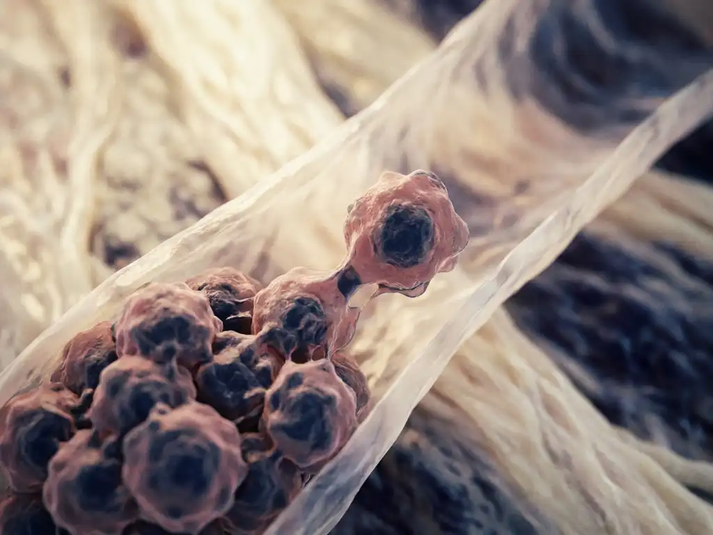
Cancer spreads when malignant cells break away from the original tumor, travel through the body via the bloodstream or lymphatic system, and establish new tumors in distant tissues. This process, known as cancer metastasis, is what makes many cancers life-threatening. The sections below unpack the key questions researchers ask about how cancer cells spread, where they go, and what science is doing to intervene.
What happens to cancer cells that makes them able to spread?
Cancer cells acquire the ability to spread through a series of biological changes that allow them to detach from their original location, invade surrounding tissue, and survive in new environments. This process is called cancer invasion, and it begins when cells lose the molecular “glue” that normally holds them in place within a tissue.
Healthy cells are anchored by proteins called adhesion molecules. In cancer, mutations can reduce the expression of these proteins, allowing cells to detach. Some cancer cells also undergo a transformation called epithelial-to-mesenchymal transition (EMT), during which they take on properties more typical of mobile, connective-tissue cells. This shift makes them physically capable of moving through dense tissue.
Additionally, cancer cells can secrete enzymes that degrade the extracellular matrix, the structural scaffold surrounding cells. By breaking down this barrier, they create pathways into blood vessels or lymphatic channels. These combined changes are what make a cancer cell metastatic rather than locally confined.
How do cancer cells travel through the body?
Cancer cells travel through the body primarily via two routes: the bloodstream (hematogenous spread) and the lymphatic system (lymphatic spread). Once a cancer cell enters either of these networks, it can be carried far from the original tumor to distant organs and tissues.
Traveling through the circulatory or lymphatic system is hostile to most cells. The majority of circulating tumor cells do not survive the journey. They face mechanical stress, immune surveillance, and a lack of the supportive signals they received in their original tissue. Only a small fraction of cells that enter the bloodstream successfully establish a new tumor elsewhere.
When cancer cells do survive, they must exit the vessel at a new site, a process called extravasation, and then adapt to an entirely different tissue environment. This adaptation phase, sometimes called colonization, is one of the most active areas of metastatic cancer research today.
Why does cancer spread to certain organs and not others?
Cancer does not spread randomly. Certain tumor types show strong preferences for specific organs, a concept researchers describe as “organotropism.” Breast cancer, for example, frequently spreads to bone, lung, liver, and brain, while prostate cancer has a particular affinity for bone. These patterns are not coincidental.
The leading explanation is the “seed and soil” hypothesis, first proposed in the 19th century and still supported by modern molecular biology. According to this model, cancer cells (the seeds) only successfully colonize organs (the soil) that provide the right molecular signals, nutrient availability, and immune environment. Organs that secrete specific growth factors or have particular surface receptors may actively attract circulating tumor cells from certain cancer types.
Research also shows that primary tumors can prepare distant organs before cancer cells even arrive by releasing signaling molecules that modify the target tissue to become more hospitable. This pre-metastatic niche formation is an important focus of current oncology research, as disrupting it could potentially prevent metastasis before it begins.
What is the difference between a primary tumor and a secondary cancer?
A primary tumor is the original site where cancer first developed. A secondary cancer, also called a metastasis or metastatic lesion, is a new tumor formed when cells from the primary tumor spread to a different part of the body. Critically, a secondary cancer retains the characteristics of the original cancer, not the organ where it has settled.
This distinction matters enormously in research and clinical contexts. For example, if breast cancer spreads to the lung, the secondary tumor in the lung is still classified as metastatic breast cancer, not lung cancer. The cells carry the molecular signature of their origin, which influences how researchers study them and which biological pathways are relevant to investigate.
Secondary cancers can develop in almost any organ and may appear months or even years after the primary tumor was first identified. In some cases, a secondary cancer is discovered before the primary tumor, prompting researchers to trace the origin using molecular profiling techniques.
What are the stages at which cancer is considered to have spread?
Cancer staging systems use a numbered scale, typically from Stage I to Stage IV, to describe how far cancer has progressed. The spread of cancer to other parts of the body is generally reflected in the higher stages of this scale.
- Stage I: The tumor is small and localized. Cancer cells have not spread beyond the original tissue.
- Stage II: The tumor has grown larger but remains largely contained. There may be limited spread to nearby lymph nodes.
- Stage III: Cancer has spread more extensively to regional lymph nodes or nearby structures, but distant organs are not yet involved.
- Stage IV: Cancer has spread to distant organs or tissues. This is the stage at which cancer is formally considered metastatic.
It is worth noting that staging criteria vary between cancer types and the classification systems used by different research and clinical frameworks. The TNM system, which evaluates tumor size, lymph node involvement, and distant metastasis, is widely used in research settings to standardize how the extent of cancer spread to organs is described and compared across studies.
Can the spread of cancer be stopped or slowed?
Research into preventing or slowing cancer metastasis is one of the most active areas in oncology today. While no approach has been shown to universally halt the spread of all cancer types, several biological mechanisms are being investigated as potential intervention points.
Key research directions include:
- Targeting EMT: Disrupting the epithelial-to-mesenchymal transition that enables cells to become mobile is being explored as a way to reduce invasiveness.
- Blocking matrix-degrading enzymes: Inhibiting the enzymes cancer cells use to break through tissue barriers may limit their ability to invade surrounding structures.
- Disrupting pre-metastatic niches: Preventing primary tumors from conditioning distant organs is an emerging strategy to stop colonization before it starts.
- Immune system engagement: Research is investigating how to enhance the body’s immune surveillance to identify and eliminate circulating tumor cells before they establish new tumors.
- Targeting organotropism signals: Understanding the molecular signals that attract cancer cells to specific organs opens the possibility of blocking those signals to redirect or prevent metastatic spread.
These approaches are being investigated for their potential to contribute to the broader goal of controlling metastatic cancer. Research in this area relies heavily on advanced laboratory tools, including imaging systems, organoid models, and high-throughput screening platforms, to study how cancer cells behave and respond to experimental interventions at the cellular and molecular level.
How BioSPX supports cancer metastasis research
Understanding how cancer spreads requires precise, reproducible laboratory tools. In the Benelux region, BioSPX supports life science researchers working on cancer biology, invasion assays, and metastasis modeling with a broad portfolio of instruments and expert guidance.
- Multimode readers and imaging systems for tracking cellular behavior and tumor cell migration
- Liquid handling solutions that improve reproducibility in high-throughput cancer research workflows
- Organoid research platforms that enable three-dimensional cancer models closer to in vivo conditions
- Automation systems that reduce manual variability in cell culture and screening experiments
- Product specialists who advise on instrument selection and experimental setup for oncology research applications
BioSPX serves academic research laboratories, biotech companies, pharmaceutical firms, and R&D organizations across Belgium, the Netherlands, and Luxembourg. If your research involves cancer cell biology or metastasis modeling, contact our team to discuss which instruments and configurations best support your work.
Frequently Asked Questions
What is the difference between cancer invasion and cancer metastasis?
Cancer invasion refers to the local spread of tumor cells into the surrounding tissue immediately adjacent to the primary tumor, while metastasis describes the full process by which cancer cells travel to and establish new tumors in distant organs or tissues. Invasion is essentially the first step that makes metastasis possible — without it, cancer cells cannot access the bloodstream or lymphatic system needed to travel. Researchers often study these two processes separately, as the molecular mechanisms driving local invasion are not always identical to those governing distant colonization.
What are circulating tumor cells (CTCs), and why do researchers study them?
Circulating tumor cells (CTCs) are cancer cells that have detached from a tumor and entered the bloodstream, making them detectable in a blood sample — a technique often referred to as a liquid biopsy. Researchers study CTCs because they represent a real-time snapshot of a patient’s metastatic potential and can provide molecular information about the cancer without requiring an invasive tissue biopsy. Analyzing CTCs allows scientists to investigate which cells survive the hostile circulatory environment, what genetic traits give them a survival advantage, and how their characteristics change over time or in response to treatment.
How do researchers model cancer metastasis in the laboratory?
Researchers use a range of experimental models to study metastasis, from two-dimensional cell migration assays (such as scratch/wound-healing assays and Transwell invasion assays) to more complex three-dimensional organoid cultures and microfluidic organ-on-a-chip systems. Animal models, particularly mouse xenograft models, are also widely used to observe metastatic behavior in a living system. Increasingly, 3D organoid platforms are preferred because they better replicate the architecture and signaling environment of real tissues, making the results more translatable to human biology.
Why is Stage IV cancer generally harder to treat than earlier-stage cancer?
At Stage IV, cancer has spread to distant organs, meaning treatment must address multiple tumor sites simultaneously rather than a single, localized mass. Metastatic tumors can also develop independently from the primary tumor over time, acquiring new mutations that may make them resistant to therapies that were initially effective. Additionally, some organs where metastases commonly form — such as the brain or bone — present unique biological barriers that limit how well certain drugs can reach and act on the tumor cells.
Can a cancer that has metastasized ever be completely eliminated?
In most cases, Stage IV metastatic cancer is considered very difficult to cure, and treatment goals often shift toward controlling the disease, extending survival, and maintaining quality of life. However, there are notable exceptions — certain cancers, such as some cases of metastatic testicular cancer or specific leukemias, can be cured even at advanced stages with the right therapeutic approach. Ongoing research into immunotherapy, targeted therapies, and combination treatment strategies is expanding the number of cancer types where long-term remission after metastasis is becoming a realistic outcome.
What common mistakes should researchers avoid when setting up cancer invasion or metastasis assays?
One of the most frequent pitfalls is poor reproducibility due to inconsistent cell seeding densities, variable gel or matrix preparation, or manual pipetting errors — all of which can skew migration and invasion results significantly. Researchers should also ensure that control conditions are rigorously maintained, including matched passage numbers for cell lines and consistent incubation environments, since even small fluctuations in temperature or CO₂ can alter cell behavior. Using liquid handling automation and validated imaging systems with standardized acquisition settings can substantially reduce inter-experiment variability and improve the reliability of results.
What should a research team consider when choosing instruments for a cancer metastasis research workflow?
Key considerations include the specific assay types your lab runs (e.g., 2D migration, 3D invasion, organoid culture, or high-throughput screening), the throughput requirements, and whether the workflow demands live-cell imaging or endpoint analysis. Compatibility between instruments — for example, between your liquid handling system and your plate reader or imaging platform — is also critical for building an efficient, reproducible pipeline. Consulting with a product specialist who understands oncology research workflows, as BioSPX offers for laboratories in the Benelux region, can help ensure that instrument selection is matched to your specific experimental goals rather than just general lab needs.
Related Articles
- What is label-free cell analysis and how does it work in oncology research?
- How do oncogenes differ from tumor suppressor genes in cancer development?
- What is liquid biopsy and how is it used in oncology?
- How does radiation therapy work in cancer treatment?
- How do oncologists decide which cancer treatment to recommend?
This content was generated with the help of AI and it may contain mistakes