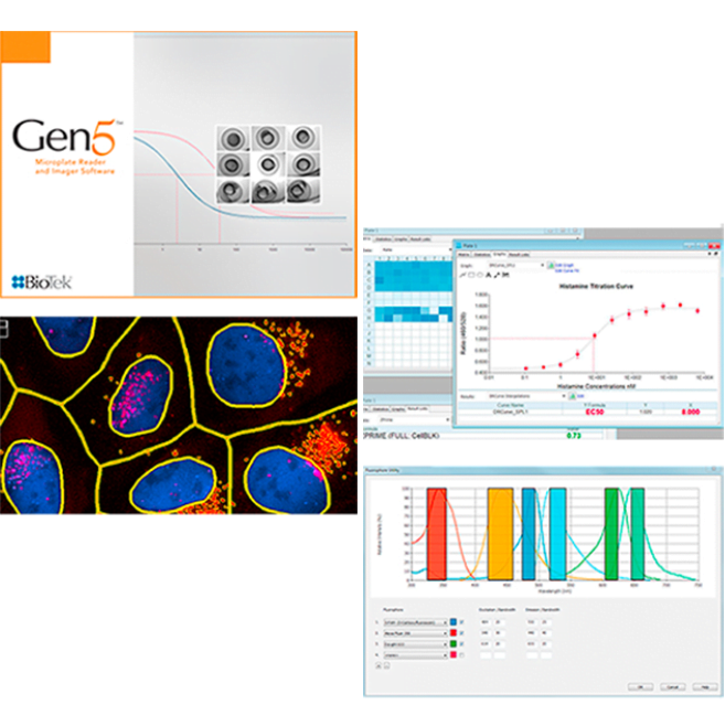
Gen5™ version 3 offers new functionality for imaging and microplate reading, including spot counting for powerful cellular image analysis and a fluorophore spectra utility for convenience during fluorescence detection protocol definition, and many others.
From high magnification, intracellular image analysis to basic ELISA, Gen5 software helps transform raw data and images into meaningful results. Gen5 Software offers a unique combination of power and ease of use that drives productivity and saves time across all imaging and detection workflows.
Imaging or scanning multiple regions of interest (ROIs) within a microplate well or slide using high magnification, such as in a slide scanning workflow, can be a time-consuming process.
The automatic Region of Interest (AutoROI) identification module available for Agilent BioTek Gen5 software uses a three-step sequence to automate the ROI identification process.
First, a low-magnification imaging step quickly images the entire area of the slide, microplate well, or another vessel. Next, ROIs are automatically identified using previously optimized criteria. Immediately following identification, high-magnification imaging is then automatically performed on the identified areas. The Auto ROI module for Gen5 software can be used with Agilent BioTek Cytation cell imaging readers and Lionheart automated microscopes. For higher-throughput slide scanning, Cytation can be integrated with Agilent BioTek BioStack microplate stacker.
Conventional image analysis relies on background flattening, object sizing, and repeated intensity thresholding to identify cells. These steps require a high level of expertise to implement and interpret, and can be difficult to apply consistently to label-free brightfield, live-cell, and kinetic images, contributing to user-to-user variability in results.
The Agilent BioTek Gen5 AI cell identification module is an add-on analysis module for BioTek Gen5 software for imaging and microscopy. It applies AI-based segmentation to identify and count cells directly, reducing the need for thresholding and parameter tuning. This supports more reliable cell counting and confluence data with less user-to-user variability, across diverse cell types and assay conditions. The result is a more efficient path from image acquisition to quantitative insight, with fewer manual analysis steps and minimal user intervention.
BioTek Gen5 Al cell identification module generates consistent image derived insights across cell types, labeling strategies, imaging magnifications, imaging conditions, and time-based experiments. 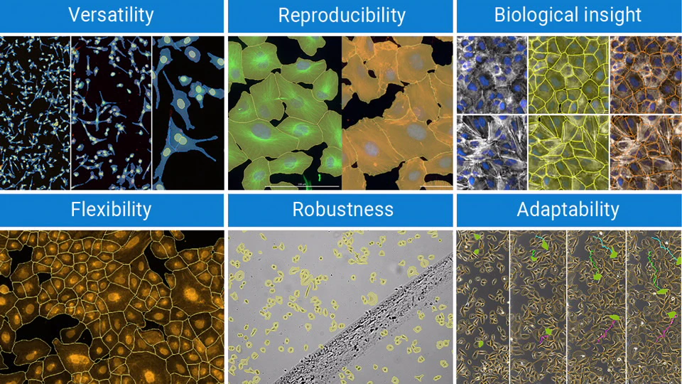
Consistent cell identification is maintained as cultures progress from sparse populations to densely packed monolayers. By adapting to changes in cell number, contact, and boundary visibility, researchers can compare results across experimental conditions with greater confidence.
Reliable segmentation is maintained when blur, debris, high background, or variable staining makes image analysis more difficult. By reducing the impact of common imaging artifacts, researchers can extract more dependable data from challenging samples and conditions.

 EN