
 EN
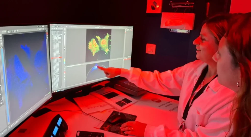
Measuring how proteins interact in their natural habitat
A deep dive into FLIM-FRET and the QF Pro® workflow
“A lot of people study protein-protein interactions by doing co-IP or mass spec on lysates,” says Eef Parthoens (VIB BioImaging Core Ghent) . “That gives you an answer, but it’s an averaged answer. What we can do with microscopy is ask: where in the tissue does this happen? In which cells? In which compartment? I think many researchers underestimate what we can do in this regard at the BioImaging Core.”
Their recent article takes a technology-focused look at Fluorescence Lifetime Imaging Microscopy (FLIM) and how it becomes a quantitative readout for protein proximity through FRET. It also zooms in on a practical workflow that has recently gained traction at VIB: QF Pro®, developed by HAWK, which combines two-site immunofluorescence with amplified FRET-FLIM to quantify protein interactions directly in fixed tissue. For the BioImaging Core, it has become both a success story and a gateway into a broader conversation about what “functional imaging” can mean in an institute that is rapidly embracing spatial biology.
Moving beyond intensity in complex samples
Instead of measuring the fluorescence intensity, FLIM measures how long a fluorophore remains in its excited state after a pulsed laser excitation. That number—the fluorescence lifetime, expressed in nano or picoseconds—is surprisingly stable and often far less sensitive to the usual sources of intensity artefacts.
“The strength of lifetime is that it’s a physical property,” Eef explains. “Instead of asking how much signal you have, you’re asking how the fluorophore behaves. That makes it much more robust when samples are challenging.”
That robustness is also why FLIM is still relatively rare in practice. It requires dedicated hardware for time-resolved detection, pulsed excitation, and analysis workflows that go beyond standard confocal imaging. “You can’t do FLIM on every confocal microscope,” Eef says. “In Belgium, there are only a handful of systems with a proper FLIM module, and you need the right setup and the expertise to make the measurements reproducible.”
Bringing quantitative interaction readouts into fixed tissue with QF Pro®
QF Pro® is a workflow designed to quantify interactions in fixed cells and tissue by combining two-site immunofluorescence with amplified FRET-FLIM. The idea is straightforward: preserve tissue architecture, label two proteins of interest, and use lifetime shifts to infer proximity in situ, even in autofluorescent tissue where conventional intensity-based approaches struggle.
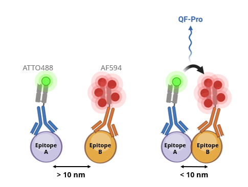
Eef first encountered the approach through BioSPX (the life science division of BRS), via Wouter De Sadeleir, who represents the technology in Belgium and the Netherlands. The initial trigger was the broader rise of spatial biology workflows at VIB.
“People are mapping tissues with more and more labels,” Eef says. “And that naturally raises the next question: not only where are proteins, but where do they interact?”
HAWK Biosystems provided test kits, and the core recruited a handful of early users. Such early pilots don’t merely test performance, it’s also a way of gauging whether the workflow is compatible with real lab timelines and scientific incentives. “It’s never only about whether the method works,” Eef notes. “Equally important is if it fits the way people actually do research.”
The evaluation has since attracted the attention of the VIB Tech Watch Core, which recognized the potential for broader institute value. Eef connected with Bram Van den Bergh to explore how to move from an early, kit-driven pilot into a more scalable evaluation path. “It’s exactly the type of technology where it helps to have a broader view,” Eef says. “You can support multiple groups, and you can also be very clear about where the method is strong and where it isn’t.”
Making functional imaging more approachable
The Ghent BioImaging Core team wants to use the QF Pro® case to make FLIM less “mystical” to the VIB research community.
“FLIM is not the easiest technique,” Eef says. “But once people understand what lifetime is and why it’s robust, they start recognizing where it can answer their questions.”
The timing is good. There is rising interest from additional groups who want to test interaction readouts in tissue, and VIB Tech Watch is exploring how to support a wider, more structured evaluation. There is also a natural connection to the institute’s broader shift toward integrated, spatial, multi-modal biology: as datasets become richer, researchers want methods that connect molecular measurements to mechanisms in context.
From the BioImaging Core perspective, the long-term value is clear. “A lot of researchers still don’t know that we can measure interactions in living cells and tissue in a quantitative way,” Eef says. “If we can make that visible, then we can help researchers turn a difficult question into a robust, quantitative assay, with the right controls and the right interpretation.”
Are you interested to read the full article? Go to https://blog.vib.be/seeing-interactions-where-biology-happens
