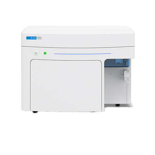
NovoCyte Advanteon
Information
Overview
The NovoCyte Advanteon flow cytometer builds upon the highly successful NovoCyte, and NovoCyte Quanteon. The system provides an advanced set of capabilities for the most demanding scientists, yet is remarkably simple to operate. The NovoCyte Advanteon can accommodate high-end and increasingly sophisticated multicolor flow cytometry assays. The system offers flexibility with 1, 2, or 3 laser options, up to 21 fluorescence channels, and 23 independent detectors.
The NovoCyte Advanteon is customizable to meet your specific needs but is easily upgradeable to meet your future demands. When throughput is essential, the NovoSampler Q can be integrated into different laboratory automation platforms. The sampler can efficiently process both FACS tubes (using a 40-tube rack) and 24-, 48-, 96-, and 384-well plates. For the NovoCyte Advanteon, the intuitive and industry honored NovoExpress software is now even more advanced, providing an exceptional user experience in data acquisition, analysis, and reporting.
• Sample recovery mode serves to collect unused sample at end of acquisition
• Excellent sensitivity and resolution
• Intuitive and powerful software for data acquisition, analysis, and reporting
• Smart-design functionalities and walk-away operation simplifies your work flow
• Automation-ready capability for high throughput needs
• Wide, 7.2-log dynamic range eliminates the need for routine gain detector adjustments
• High speed collection up to 100,000 events/second
• Accurate absolute cell count in every experiment, which eliminates the need for reference beads
-
Models
Product Lasers 349 nm 405 nm 488 nm 561 nm 637 nm Maximum Number of Fluorescence Channels NovoCyte Advanteon** 1 • 7 • 6 2 • • 15 • • 13 • • 11 3 • • • 21 • • • 17 • • 19 NovoCyte Quanteon** 4 • • • • 25 NovoCyte Penteon* 5 • • • • • 30 * RUO: Research use only. Not for use in diagnostic procedures. ** Selected configurations are registered as CE-IVD. -
Applications
Apoptosis Assay
Apoptosis, or programmed cell death, is the process by which cells regulate how they die, activating specific pathways that cause the cell to shrink, condense, and eventually be cleared by phagocytosis. This is in contrast to necrotic cell death where cells die uncontrollably and fall apart, which can lead to detrimental effects such as the activation of an immune response. Therefore, apoptotic cells that die in a very orderly fashion limit disruption of nearby cells and tissue.
There are many ways to measure cell death and distinguish it from apoptosis or necrosis. These assays are easily quantified using the NovoCyte flow cytometer due to automatic compensation settings and a wide dynamic range of fluorescence detection which eliminates the need for any PMT voltage adjustments.Immunophenotyping
Immune status is associated with disease state, treatment efficiency, and response to external stimuli such as vaccines. Immunophenotyping quickly identifies candidate cell types, sub-classes and functions. Monitoring the frequency of numerous immune cell population as well as the differentiation/activation status of specific cell subsets such as monocytes, NK cells, T and B cells is essential as they may influence the immunogenicity of a vaccine and its efficiency. The NovoCyte Flow Cytometer enables simultaneous quantification of multiple leukocytes for better understanding the immune status of patients and surveillance of the immune response to infectious disease.Specifcity Clone Fluorocrome Purpose CD3 UCHT1 PE-TR (ECD) Lineage T cells CD4 S3.6 PE-Alexa 700 Lineage T cells CD8 SK1 PerCP-Cy5.5″ Lineage T cells CD19 J3-129 PerCP-eFluor 710 B cells CD14 MφP9 BV711 Monocytes CD56 HCD56 BV605 NK cells and NK T-like cells CD16 3G8 APC-Cy7 NK cells and monocytes γδ TCR 11F2 PE-Cy7 γδ T cells Vγ2 TCR B6 PE γδ T cells CD25 M-A251 BV421 Tregs CD127 A019D5 APC Tregs/memory/differentiation CD45RA HI100 BV650 Memory/differentiation CCR7 G043H7 BV785 Memory/differentiation CD57 NK-1 FITC Memory/differentiation HLA-DR B169414 BV570 Activation CD38 HIT2 PE-Cy5 Activation/plasmablasts NKG2C 134591 Alexa 700 NK receptor Dead Cells 134591 AViD Dead cell exclusion Intracellular Protein Detection
Detection and analysis of intracellular proteins allow for additional characterization of cell subpopulations and cellular processes. In order to analyze proteins not located on the cell surface, fixation and permeabilization of the cell is required. However, many phospho-specific antibodies are not compatible with many common detergent-based permeabilization methods used for intracellular staining. Special attention is needed when determining the proper fix/perm method for your phospho-specific antibody. The most common method uses 1.5% paraformaldehyde for fixation followed by 100% methanol for permeabilization. While this method works for many antibodies, please note it may not work for every phospho-specific antibody.Additionally, identifying various cell populations in a heterogenous sample requires staining for phosphorylated proteins coupled with surface proteins. Special consideration must be given to the sensitivity of these epitopes to fixative, taking precaution to avoid damage to the epitope. Therefore, the sample may require staining for specific surface markers before fixation.
Cell Cycle Analysis
Normal human somatic cells are diploids containing a constant amount of DNA. During cell cycle progression, DNA synthesis results in a doubling of total DNA content, followed by restoration of the normal DNA content after mitosis. Detailed cell cycle analysis can be performed to understand tumor cell differentiation, cell transformation and cell-compound interaction with the NovoCyte flow cytometer.Figure: After treatment with 10 migrograms/M MG132 or 500 micrograms/M 5-FI for 16 hours/ A549 cells were analyzed for cell cycle distribution with the ACEA Novocyte flow cytometer. The the Novoexpress built-in cell cycle analysis module, the plot shows cells ni G0/G1 phase (green), S phase (yellow) and G2/M phase (blue). Compared to normal untreated cells, MG132 treated cells were arrested at G2/M phase, while 5-FU treated cells were arrested at G0/G1 phase.
Cell Proliferation
Cell proliferation is an essential function and highly structured event that when unregulated, can cause disease. We can measure proliferation through absolute cell counts or with a dye, such as CFSE. When cells labeled with CFSE divide, the dye is partitioned equally between daughter cells and we can measure the loss of CFSE fluorescence over time as the dye is continuously diluted. The mean fluorescence intensity (MFI) of the dye was also plotted with cell concentration over time to show the inverse relationship between the two. This type of assay is often used to look at changes in T lymphocyte activation. -
How it works
The Ultimate Photodetector
Silicon photomultipliers (SiPM) are solid-state, silicon-substrate-based, photon-level-sensitive semiconductor devices, with a 7.2 log dynamic range. Consisting of a compact array of avalanche photodiodes operating in unison, the SiPM is a compact detector with photon counting capability. The innovative optics designed into the NovoCyte Penteon incorporates 30 independent SiPM, which collect and process signals for each of its fluorescence channels.Excellent scatter resolution to detect small particles
NovoCyte Penteon scatter detection optics and signal processing electronics have been optimized to resolve particles down to 0.1μm in size. With such excellent resolution, platelets, bacteria, and various submicron particles can be readily identified and analyzed.Consistent results, fast or slow
The fluidic feedback control mechanisms found on the NovoCyte Advanteon consistently maintain exceptionally steady flow rates. The superior stability across a wide range of sample flow rates provides consistent results under variable operating conditions. As shown here, the CVs at different flow rates are dramatically improved over other systems on the market. -
Features
General
The NovoCyte Advanteon is an easy to handle and flexible instrument that provides multicolour flow cytometry assays. The system has 1-3 laser options and up to 21 fluorescence channels, plus forward and side scatter. The silicon photomultipliers (SiPM) in the Advanteon deliver photon-level sensitivity.The high sensitivity and resolution instrument is able to operate with FACS tubes and 24-, 48-, 96-, 384-well plates. For the NovoCyte Advanteon, the intuitive NovoExpress software is now even more advanced, providing an exceptional user experience in data acquisition, analysis and reporting.
Brochures
Application notes
Please login to be able to see the application notes.

