
 EN
Beyond Co-localisation: Measuring Protein–Protein Interactions in FFPE Tissue

The core question spatial biology has not yet answered...
Are two proteins actually interacting? Right here, in this cell, in this patient’s tissue? Not co-expressed. Not adjacent.
Interacting! QF-Pro® from HAWK Biosystems is the first technology to answer that question reliably, quantitatively, and reproducibly in FFPE clinical samples.
1 – 10 nm
Interaction resolution Actual molecular binding scaleFFPE
Works in archival formalin-fixed paraffin-embedded patient tissue
Quantitative
Robust interaction score reproducible across cohorts
Single-cell
Spatial resolution — cell-by-cell mapping across tissue sections
Table of Contents
The gap that spatial biology has not yet filled
Modern spatial biology platforms have transformed how researchers read tissue.
- Transcriptomic methods reveal which genes are active in which cells;
- Multiplex immunofluorescence identifies dozens of proteins simultaneously;
- Digital pathology tools extract morphological features at scale.
Together, these technologies have given researchers an extraordinarily detailed view of tissue architecture and cell identity.
But cellular function is not determined by what proteins are present, or even how much of each is expressed. Function is determined by what those proteins do — which means who they interact with, whether those interactions are occurring, and at what intensity. A kinase that is abundantly expressed but not phosphorylating its substrate is functionally silent. A checkpoint receptor that is highly expressed but not engaging its ligand is not suppressing T cell activity. Expression tells you the cast. Interaction tells you whether the play is actually running.
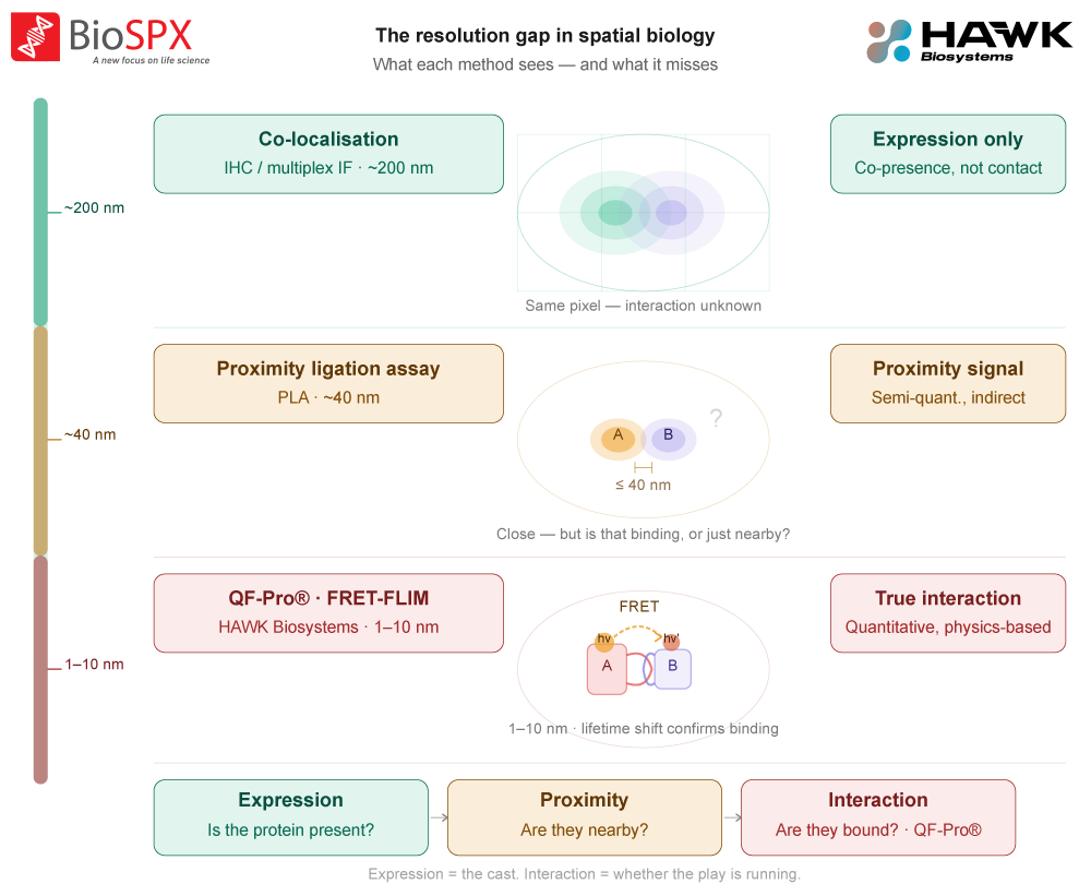
This is the gap that existing spatial biology methods have not been able to fill. Proteins interact at nanometre scales.
Light microscopy resolves at ~200 nm. Co-localisation experiments tell you that two proteins are in the same pixel, a distance potentially a hundred times larger than the binding site itself. Proximity ligation assays (PLA) detect proteins within ~40 nm, which is closer but still a probabilistic signal rather than a direct measurement of molecular engagement. Neither provides a true quantitative readout of interaction state.
What QF-Pro® actually measures, and how
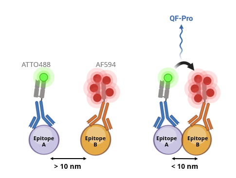
QF-Pro® (Quantifying Functions in Proteins) from HAWK Biosystems measures Förster Resonance Energy Transfer (FRET), a quantum mechanical phenomenon in which energy is transferred directly between two fluorophores when they are within 1–10 nm of each other. This distance range corresponds to actual molecular contact: if FRET occurs, the two labelled epitopes are genuinely interacting.
FRET has been understood for decades. The challenge has always been making it work reliably in complex, autofluorescent, fixed patient tissue. HAWK Biosystems has solved this with a patented signal amplification step that boosts FRET efficiency to levels measurable above the noise of FFPE samples — for the first time.
The readout is captured using Fluorescence Lifetime Imaging Microscopy (FLIM), which measures how long a fluorophore remains in its excited state before emitting a photon. When FRET occurs — when the two proteins are truly interacting — the donor fluorophore’s lifetime shortens in a measurable, quantifiable way. This lifetime shift is physics-based, not threshold-based, which is why the resulting score is genuinely quantitative and reproducible across different labs, instruments, and cohorts.
How it works, step by step
| 1 | Two-site immunolabelling Two epitopes are labelled simultaneously using species-distinct antibodies, each conjugated to a FRET-compatible donor or acceptor fluorophore. The workflow is analogous to standard immunofluorescence. |
| 2 | Patented amplification step HAWK’s proprietary amplification chemistry boosts the FRET signal to levels reliably detectable above the autofluorescence background of FFPE tissue — the key innovation that makes quantitative FRET in clinical samples possible for the first time. |
| 3 | FLIM-based interaction quantification The Violet 3.0 platform measures fluorescence lifetime pixel by pixel across the tissue section. Where FRET occurs — where the two proteins are within 1–10 nm — the donor lifetime shortens. The magnitude of this shift gives a direct, quantitative measure of interaction state. |
| 4 | Spatial quantitative score Software automatically maps interaction states across the tissue at single-cell resolution. The output is a robust, reproducible quantitative score that captures the heterogeneity of protein interaction within the tumour microenvironment. |
QF-Pro® vs. existing spatial biology approaches
Method | Resolution | True interaction | FFPE compatible | Quant. score | Single-cell spatial |
Co-localisation (mIF/IHC) | ~200 nm | No | Yes | No | Yes |
Proximity Ligation Assay (PLA) | ~40 nm | Proximity only | Limited | Semi-quant. | Yes |
Co-immunoprecipitation (Co-IP) | Molecular | Yes | No | Bulk only | No |
Standard FRET/FLIM | 1-10 nm | Yes | Not reliably | Yes | Yes |
QF-Pro® (HAWK Biosystems) | 1-10 nm | Yes | Yes – validated | Yes – robust score | Yes |
QF-Pro® operates in a measurement space that existing methods cannot access. The table below shows where each technology sits.

Clinical validation: PD-1/PD-L1 engagement predicts immunotherapy response
The most compelling evidence for what QF-Pro® unlocks comes from immuno-oncology. Only 12–20% of solid tumour patients respond to PD-1/PD-L1 checkpoint inhibitors, and PD-L1 expression by IHC is an unreliable predictor. The reason is fundamental: expressing PD-L1 does not tell you whether PD-1 and PD-L1 are actually engaging.
Journal of Clinical Oncology, 2023 — 188 NSCLC patients
In a study published in the Journal of Clinical Oncology (doi: 10.1200/JCO.22.01748), QF-Pro® was used to quantify PD-1/PD-L1 interaction state in outcome-blinded FFPE tissue from 188 patients with non-small cell lung cancer (NSCLC) who subsequently received immune checkpoint inhibitor therapy.
PD-1/PD-L1 interaction state measured by QF-Pro® was highly predictive of both progression-free survival and overall survival — outcomes that PD-L1 expression TPS by IHC failed to predict. Critically, 22.5% of patients with high PD-L1 expression but low QF-Pro® interaction scores did not respond to therapy.
Source: Calleja et al., Journal of Clinical Oncology, 2023. doi: 10.1200/JCO.22.01748
This result illustrates the core argument for functional spatial biology: the difference between expressing a protein and using it is clinically meaningful. QF-Pro® makes that difference measurable for the first time in routine FFPE tissue.
Validated assay targets
QF-Pro® is validated across a growing panel of clinically relevant protein–protein interactions and post-translational modifications, spanning immune checkpoint biology, oncogenic signalling, and activation state readouts.
Immune Checkpoint Interaction PD-1 / PD-L1 Validated in NSCLC, ccRCC, melanoma, CRC, tonsil. Published clinical data (JCO 2023). | Immune Checkpoint Interaction CTLA-4 / CD80 Validated in cell models, NSCLC, CRC-derived lung metastases, tonsil. |
Immune Checkpoint Interaction TIGIT / CD155 Validated in cell models, NSCLC, melanoma, tonsil. | Immune Checkpoint Interaction LAG-3 / MHC-II Validated in NSCLC, lymph nodes, thymus, appendix. |
Immune Checkpoint Interaction TIM-3 / Galectin-9 Validated in NSCLC and tonsil. | Oncogenic Signalling HER2 / HER3 dimerisation Validated in cell models and FFPE tissue. Relevant for HER2+ cancer target engagement. |
Activation State (PTM) Akt activation state Validated in HNSCC, ccRCC, breast, prostate, sarcoma. Phosphorylation-state readout. | Activation State (PTM) STAT3 activation state Validated in ccRCC and cell models. Y705 and S727 phosphorylation sites. |
Beyond validated assays, QF-Pro® can be applied to any protein pair for which suitable antibodies exist. HAWK Biosystems offers a custom assay development service for academic or industrial partners seeking novel interaction targets.
HAWK Biosystems
Violet 3.0 is the dedicated imaging platform for QF-Pro® assays. Designed to make FLIM-FRET accessible to any laboratory — not just specialist physics groups — it integrates high-performance fluorescence lifetime detection, automated image analysis, and an intuitive graphical interface in a single benchtop instrument.
Compatible with both standard tissue slides and multi-well plates for cell-based assays. QF-Pro® software handles all FRET-FLIM calculation automatically — no manual curve fitting, no specialist optics expertise required.

Violet 3.0
FAQ
QF-Pro® measures actual protein–protein interactions at 1–10 nm resolution — the scale of molecular binding — using a patented FRET-FLIM approach validated for FFPE patient tissue. Proximity ligation assays detect proteins within ~40 nm, which is proximity, not interaction. Co-localisation by immunofluorescence works at ~200 nm, far larger than any molecular binding event. QF-Pro® delivers a robust quantitative score rather than a binary or semi-quantitative signal.
Yes. QF-Pro® is specifically designed and validated for FFPE tissue — the standard format of clinical biobanks and pathology archives. It can be applied to existing sample collections without requiring fresh or frozen material, making retrospective cohort studies significantly more accessible.
The QF-Pro® readout is based on fluorescence lifetime, a physics-based measurement that is inherently calibrated and independent of fluorophore concentration or illumination variability. This makes the resulting interaction score directly comparable across different samples, batches, and laboratories, a critical requirement for clinical biomarker studies and multi-site trials.
BioSPX is the exclusive distributor of Violet 3.0 and QF-Pro® reagent kits in Belgium, the Netherlands, and Luxembourg. Our team can support you from first contact through instrument demonstration, installation, and training.